 Indietro
Introduction to Cell Biology: Cell Theory, Structure, and Diversity
Guida di studio - Note intelligenti
 Appunti personalizzati basati sui tuoi materiali, ampliati con definizioni chiave, esempi e contesto.
Overview of Cell Biology
Cell Theory
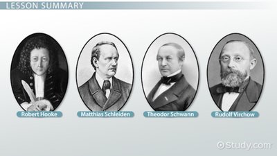
The development of cell theory was a foundational event in biology, formulated by the combined work of Schleiden, Schwann, and Virchow, enabled by advances in microscopy initiated by Robert Hooke and others. The basic tenets of cell theory are:
All organisms are composed of one or more cells.
The cell is the basic unit of structure, function, and reproduction in organisms.
All cells arise from preexisting cells.
Cells are systems of integrated molecules and organelles, and the properties of life are emergent properties of this system. In unicellular organisms, a single cell performs all life functions, while in multicellular organisms, specialized cells cooperate and become interdependent, forming higher levels of biological organization.
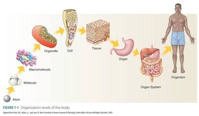
Chemical Components and Structure of Cells
Cellular Organization and Diversity
Cells exhibit a wide diversity in structure, which is closely related to their function. The cell is a living unit greater than the sum of its parts, with emergent properties arising from the integration of its molecular components.
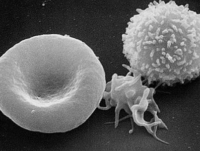
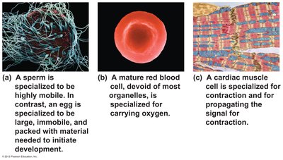
Examples of specialized cells include:
Sperm cell: Small with a tail for motility, specialized for fertilization.
Red blood cell: Biconcave shape, filled with hemoglobin, specialized for oxygen transport.
Muscle cell: Cylindrical and packed with contractile filaments, specialized for contraction.
Techniques in Cell Biology
Microscopy
Microscopy is essential for studying cells. Two main types are:
Light Microscope: Magnification up to 1,000X; resolution 200–350 nm.
Electron Microscope: Magnification up to 100,000X; resolution 0.2–2 nm. Allows for 3D imaging and visualization of ultrastructure.
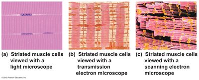
Basic Cell Structure
Universal Features of Cells
All cells share several fundamental structures:
Plasma Membrane: Lipid bilayer that encloses the cell's contents.
Chromosomes: Units of genetic material (DNA).
Cytoplasm: Cytosol and suspended structures.
Ribosomes: Sites of protein synthesis.
Cytoskeleton: Protein framework for support, shape, and movement.
Prokaryotic and Eukaryotic Cells
Major Cell Types
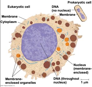
Cells are classified into two basic types:
Prokaryotic Cells: Bacteria and Archaea; lack a nucleus and most organelles.
Eukaryotic Cells: Animals, plants, fungi, and protists; possess a nucleus and membrane-bound organelles.
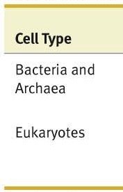
Cell Type | Location of DNA | Internal Membranes and Organelles |
|---|---|---|
Bacteria and Archaea | In nucleoid (not membrane bound); plasmids also common | Extensive internal membranes only in photosynthetic species; limited types and numbers of organelles |
Eukaryotes | Inside nucleus (membrane bound); plasmids extremely rare | Large numbers of organelles; many types of organelles |
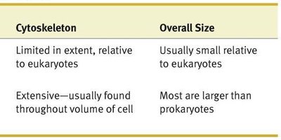
Cytoskeleton | Overall Size |
|---|---|
Limited in extent, relative to eukaryotes | Usually small relative to eukaryotes |
Extensive—usually found throughout volume of cell | Most are larger than prokaryotes |
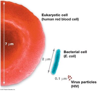
Relative sizes of cells and viruses:
Viruses: Acellular Infectious Agents
General Features of Viruses
Viruses are extremely small, infectious, acellular particles. They are obligate intracellular parasites, requiring host cell machinery for reproduction and protein synthesis. Viruses infect specific host species and cell types, and are the most abundant biological entities on Earth.

Viruses lack cellular organization, independent metabolism, and reproduction.
They possess genetic material and can evolve by natural selection.
Viruses have contributed significantly to our understanding of cell biology and molecular genetics.
Structure of Viruses
Nucleic acid genome (DNA or RNA, single- or double-stranded)
Protective protein capsid (composed of repeating protein subunits)
Some have a membranous envelope derived from the host cell
Capsid morphology can be helical, icosahedral, or complex. The envelope contains viral glycoproteins and host membrane components.
Viral Life Cycle (Generalized Stages)
Attachment to host cell
Entry of viral genome
Genome replication
Protein synthesis
Assembly of new virus particles
Release from the host cell
Eukaryotic Cell Structure and Function
Nucleus
The nucleus is the information center of the cell, housing most genetic material in the form of chromosomes. It regulates gene expression, synthesizes rRNA, and assembles ribosomal subunits in the nucleolus. The nuclear envelope is a double membrane with nuclear pores for molecular transport. Chromatin consists of DNA and histone proteins, while the nuclear matrix organizes chromatin and supports nuclear functions.
Ribosomes
Ribosomes are non-membranous organelles composed of rRNA and proteins. They carry out translation (protein synthesis) either free in the cytosol or bound to the rough endoplasmic reticulum (rER). Each ribosome consists of a large and a small subunit.
Cytoskeleton
The cytoskeleton is a dynamic network of protein fibers that provides mechanical support, maintains cell shape, and enables movement. It interacts with motor proteins for intracellular transport and cell motility. Examples include actin microfilaments, which support structures like microvilli and neuronal processes.
Mitochondria and Chloroplasts
Mitochondria are the sites of cellular respiration, generating ATP from nutrients. Chloroplasts are the sites of photosynthesis, converting light energy into chemical energy. Both organelles are surrounded by a double membrane and contain their own DNA and ribosomes, supporting the endosymbiont theory of their origin.
Mitochondria: Inner membrane forms cristae to increase surface area for respiration.
Chloroplasts: Internal membrane system houses photosynthetic machinery.
Other Organelles
Peroxisomes: Break down fatty acids and detoxify substances using catalase.
Lysosomes: Contain digestive enzymes for breaking down macromolecules and recycling cellular components.
Vacuoles: Prominent in plant cells for storage, support, and waste degradation; contractile vacuoles expel excess water in protists.
Endomembrane System
Components and Functions
The endomembrane system synthesizes, modifies, sorts, and transports proteins and lipids. It includes:
Endoplasmic Reticulum (ER): Smooth ER (lipid synthesis, detoxification, Ca2+ storage); Rough ER (protein synthesis and modification).
Golgi Apparatus: Modifies, sorts, and packages proteins and lipids for delivery.
Transport Vesicles: Shuttle cargo between organelles and to the plasma membrane.
Lysosomes, Vacuoles, Plasma Membrane: Associated with the system for digestion, storage, and secretion.
Extracellular Structures
Cell Walls and Extracellular Matrix
Many cells synthesize and secrete extracellular structures for support, protection, and communication. Plant, bacterial, archaeal, fungal, and some protist cells have cell walls, which maintain shape, protect against stress, and join cells into tissues. Plant cell walls have three layers: primary cell wall, secondary cell wall, and middle lamella.
Form Fits Function
Specialization of Cells
The size, shape, and composition of a cell are closely correlated with its function. For example, pancreatic cells are packed with rough ER and Golgi for enzyme secretion, testis cells with smooth ER for steroid synthesis, leaf cells with chloroplasts for photosynthesis, and cardiac muscle cells with mitochondria for energy production.
