 Indietro
Membrane Structure and Function: The Fluid Mosaic Model and Its Components
Guida di studio - Note intelligenti
 Appunti personalizzati basati sui tuoi materiali, ampliati con definizioni chiave, esempi e contesto.
Membrane Structure and Function
The Functions of Biological Membranes
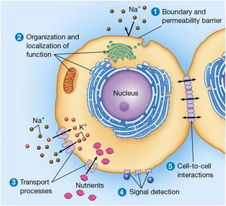
Biological membranes are essential for cellular life, serving multiple distinct roles that are fundamental to cell biology. These functions include boundary definition, compartmentalization, transport regulation, signal detection, and cell-to-cell communication.
Boundary and Permeability Barrier: Membranes define the boundaries of cells and organelles, acting as selective barriers that regulate the passage of substances.
Organization and Localization of Function: Membranes provide sites for specific biochemical activities, such as electron transport in mitochondria and protein processing in the endoplasmic reticulum.
Transport Processes: Membrane proteins facilitate and regulate the movement of ions, nutrients, and other molecules across membranes.
Signal Detection: Membrane proteins act as receptors, detecting extracellular signals and initiating cellular responses.
Cell-to-Cell Interactions: Membranes mediate adhesion and communication between cells, crucial for tissue formation and function.
Additional info: These functions are directly related to the chemical composition and structural features of membranes, which are discussed in detail in subsequent sections.
Models of Membrane Structure: Historical Perspective
Development of Membrane Models
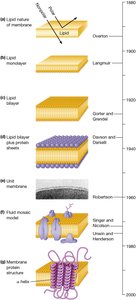
The understanding of membrane structure has evolved through a series of experimental discoveries, culminating in the fluid mosaic model. This model describes membranes as dynamic, fluid bilayers of phospholipids with proteins embedded or attached, forming a mosaic of functional components.
Overton (1890s): Proposed the presence of lipids on the cell surface based on permeability studies.
Langmuir (1910s): Demonstrated phospholipid monolayers, showing amphipathic orientation.
Gorter and Grendel (1925): Established the lipid bilayer as the basic membrane structure.
Davson and Danielli (1935): Introduced the protein-lipid-protein "sandwich" model.
Robertson (1950s): Proposed the unit membrane concept based on electron microscopy.
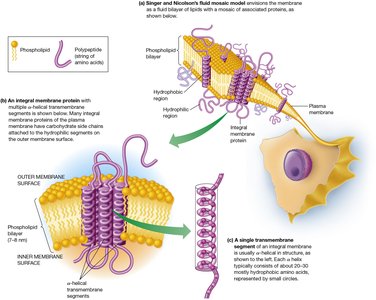
Singer and Nicolson (1972): Developed the fluid mosaic model, recognizing proteins as discrete entities within a fluid lipid bilayer.
Unwin and Henderson (1970s): Revealed transmembrane segments in membrane proteins.
Additional info: The fluid mosaic model remains foundational, though recent research highlights the importance of lipid and protein microdomains for specialized functions.
Experimental Evidence for Membrane Structure
Electron Microscopy and Membrane Visualization
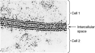
Electron microscopy provided direct visual evidence for the existence and structure of cellular membranes. Staining techniques revealed the trilaminar appearance of membranes, supporting the bilayer concept.
Trilaminar Pattern: Membranes appear as two parallel dark lines separated by a lighter zone, corresponding to the hydrophilic head groups and hydrophobic interior.
Unit Membrane: All cellular membranes share a common underlying structure.
Protein and Lipid Content of Biological Membranes
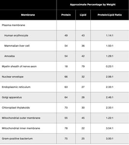
Membranes vary in their protein-to-lipid ratios, reflecting their specialized functions. For example, the inner mitochondrial membrane is protein-rich due to its role in electron transport and ATP synthesis.
Membrane | Protein (%) | Lipid (%) | Protein/Lipid Ratio |
|---|---|---|---|
Human erythrocyte | 49 | 43 | 1.14:1 |
Mammalian liver cell | 54 | 36 | 1.50:1 |
Amoeba | 58 | 42 | 1.29:1 |
Myelin sheath of nerve axon | 18 | 79 | 0.23:1 |
Nuclear envelope | 66 | 32 | 2.06:1 |
Endoplasmic reticulum | 63 | 27 | 2.33:1 |
Golgi apparatus | 64 | 30 | 2.46:1 |
Chloroplast thylakoids | 68 | 29 | 2.33:1 |
Mitochondrial outer membrane | 55 | 45 | 1.22:1 |
Mitochondrial inner membrane | 78 | 22 | 3.54:1 |
Gram positive bacterium | 75 | 25 | 3.00:1 |
The Fluid Mosaic Model: Structure and Features
Key Features of the Fluid Mosaic Model
The fluid mosaic model describes membranes as fluid bilayers of phospholipids with proteins embedded or attached. Lipids and proteins can move laterally, allowing dynamic interactions and functional specialization.
Lipid Bilayer: Provides a hydrophobic barrier and structural foundation.
Integral and Peripheral Proteins: Integral proteins span or are embedded in the bilayer; peripheral proteins are attached to membrane surfaces.
Protein Mobility: Most proteins and lipids exhibit lateral mobility, though some are anchored to cytoskeletal elements.
Membrane Lipids: Classes and Properties
Major Classes of Membrane Lipids
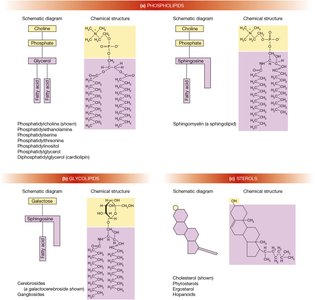
Membranes contain three main classes of lipids: phospholipids, glycolipids, and sterols. Each class contributes to membrane structure and function.
Phospholipids: Amphipathic molecules with a polar head and two nonpolar tails; form the basic bilayer structure.
Glycolipids: Lipids with attached carbohydrate groups; important for cell recognition and signaling.
Sterols: Multi-ring molecules (e.g., cholesterol) that modulate membrane fluidity and stability.
Phospholipid Composition in Different Membranes
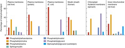
The types and proportions of phospholipids vary among membranes from different sources, affecting their properties and functions.
Fatty Acids in Membrane Lipids
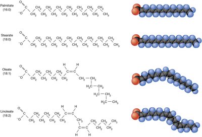
Fatty acids are essential for membrane structure, forming the hydrophobic barrier. They vary in chain length and degree of saturation, influencing membrane fluidity.
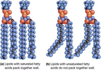
Saturated Fatty Acids: No double bonds; pack tightly, making membranes less fluid.
Unsaturated Fatty Acids: One or more double bonds; introduce kinks, increasing fluidity.
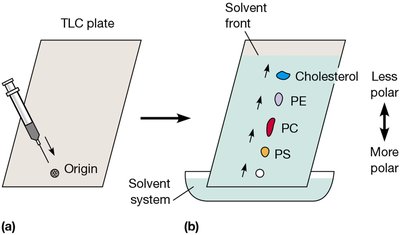
Thin-Layer Chromatography (TLC) for Lipid Analysis
TLC is used to separate membrane lipids based on polarity, aiding in the identification and study of lipid components.
Membrane Asymmetry and Lipid Movement
Asymmetry of Lipid Distribution
Membrane lipids are distributed unequally between the two monolayers, contributing to functional specialization and signaling.
Outer Monolayer: Enriched in glycolipids and phosphatidylcholine.
Inner Monolayer: Enriched in phosphatidylethanolamine, phosphatidylinositol, and phosphatidylserine.
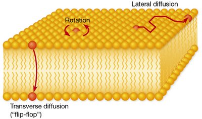
Types of Lipid Movement
Phospholipids exhibit three types of movement within membranes: rotation, lateral diffusion, and transverse diffusion (flip-flop).
Membrane Fluidity: Factors and Regulation
Transition Temperature and Membrane Fluidity
Membrane fluidity is temperature-dependent, with a characteristic transition temperature () marking the gel-to-fluid phase change. Membranes must remain fluid for proper function.
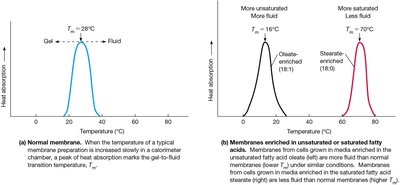
Effects of Fatty Acid Chain Length and Unsaturation
Longer fatty acid chains and higher saturation increase transition temperature and decrease fluidity. Unsaturated fatty acids lower and increase fluidity.
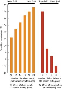
Cis vs. Trans Double Bonds
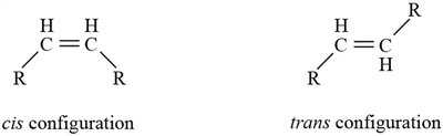
Cis double bonds introduce bends in fatty acid chains, increasing fluidity. Trans double bonds allow tighter packing, resembling saturated fats and decreasing fluidity.
Role of Sterols in Membrane Fluidity
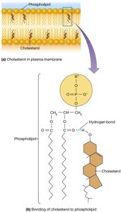
Sterols, such as cholesterol, modulate membrane fluidity by preventing tight packing of phospholipids at low temperatures and reducing fluidity at high temperatures. They also decrease membrane permeability to ions and small molecules.
Membrane Proteins: Types and Functions
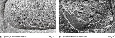
Freeze-Fracture Microscopy and Protein Mosaic
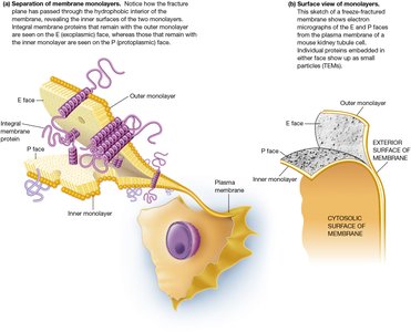
Freeze-fracture electron microscopy reveals proteins embedded within the lipid bilayer, supporting the mosaic concept of the fluid mosaic model.
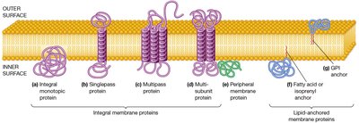
Classes of Membrane Proteins
Membrane proteins are classified based on their association with the lipid bilayer:
Integral Membrane Proteins: Embedded within the bilayer; include monotopic, singlepass, multipass, and multisubunit proteins.
Peripheral Membrane Proteins: Attached to membrane surfaces via noncovalent interactions.
Lipid-Anchored Proteins: Covalently attached to lipid molecules within the bilayer.
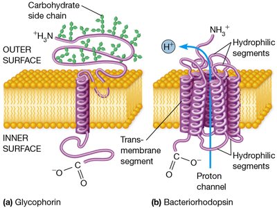
Structures of Integral Membrane Proteins
Integral proteins may span the membrane once or multiple times, often as α-helical segments. Examples include glycophorin (singlepass) and bacteriorhodopsin (multipass).
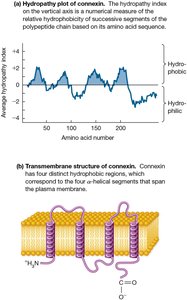
Hydropathy Analysis for Transmembrane Segments
Hydropathy plots predict the number and location of transmembrane segments based on amino acid hydrophobicity.
Membrane Protein Orientation and Glycosylation
Asymmetry of Protein Orientation
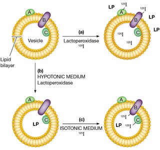
Membrane proteins are oriented asymmetrically, with specific regions exposed on either the inner or outer membrane surface. Labeling techniques can distinguish protein orientation.
Glycosylation of Membrane Proteins
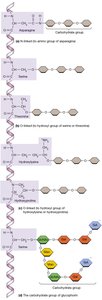
Many membrane proteins are glycosylated, with carbohydrate chains attached via N-linked or O-linked glycosylation. Glycoproteins play key roles in cell recognition and adhesion.
Membrane Protein Mobility and Structural Networks
Mobility of Membrane Proteins
Membrane proteins vary in their mobility, with some diffusing freely and others restricted by anchoring to cytoskeletal or extracellular structures. Experimental evidence from cell fusion and fluorescence recovery after photobleaching demonstrates protein mobility and domain formation.
Structural Features of the Erythrocyte Plasma Membrane
The erythrocyte membrane contains a meshwork of peripheral and integral proteins, such as spectrin, ankyrin, and glycophorin, providing mechanical support and maintaining cell shape.
Additional info: The molecular basis of membrane structure and function is central to understanding cellular processes, including transport, signaling, and cell communication. The fluid mosaic model integrates historical and experimental evidence, providing a framework for ongoing research in cell biology.
