 Indietro
Microscopy and Cell Morphology: Laboratory Foundations in Cell Biology
Guida di studio - Note intelligenti
 Appunti personalizzati basati sui tuoi materiali, ampliati con definizioni chiave, esempi e contesto.
The Light Microscope: Structure and Function
Introduction to Light Microscopy
The light microscope is a fundamental tool in cell biology, allowing for the detailed observation and measurement of structures too small for the naked eye. It utilizes visible light and a system of lenses to magnify specimens, making it essential for studying cell morphology and structure.
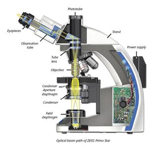
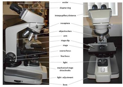
Microscope Components: The microscope consists of mechanical and optical parts. The optical system includes the condenser, objective, and ocular lenses.
Condenser: Focuses light onto the specimen, enhancing image quality but not magnification.
Objective Lens: Magnifies the specimen and projects the image toward the ocular lens.
Ocular Lens: Further magnifies the image for viewing by the eye or camera.
Total Magnification: Calculated by multiplying the magnification of the objective and ocular lenses.
Resolution and Numerical Aperture
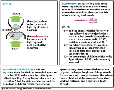
Resolution is the ability of a microscope to distinguish two points as separate entities. It is a critical factor in image clarity and detail, independent of magnification. The resolving power depends mainly on the objective lens and is defined by the following equation:
λ (lambda): Wavelength of light used (commonly 0.53 μm for white light).
n: Refractive index of the medium between the specimen and the objective lens.
θ (theta): Half the angular width of the cone of light entering the objective lens.
Numerical Aperture (NA): Defined as , it reflects the light-gathering ability of the lens. Higher NA yields better resolution and brightness.
Proper Use and Handling of the Microscope
Correct handling and focusing techniques are essential for both the safety of the instrument and the quality of observations.
Coarse Adjustment: Use only with the 4x objective to avoid damaging slides or lenses.
Fine Adjustment: Used with higher power objectives (10x, 40x, 100x) for precise focusing.
Cleaning: Only lens paper and appropriate cleaner should be used on optical surfaces.

Step-by-Step Procedure for Viewing Specimens
Following a systematic approach ensures optimal visualization and prevents damage to both the specimen and the microscope.
Clean slides and coverslips with lens paper.
Set the microscope to the lowest power (4x) and maximize working distance.
Place and center the slide on the stage.
Turn on the light and adjust the condenser and diaphragm for optimal illumination.
Focus first with the coarse, then fine adjustment knobs.
Switch to higher magnifications as needed, using only the fine adjustment knob.
After observation, clean all lenses and turn off the microscope.
Cell Morphology: Forms and Varieties of Cells
Overview of Cell Types
Cells in the human body exhibit a wide range of sizes, shapes, and functions. Their morphology is closely related to their specific roles within tissues and organs.
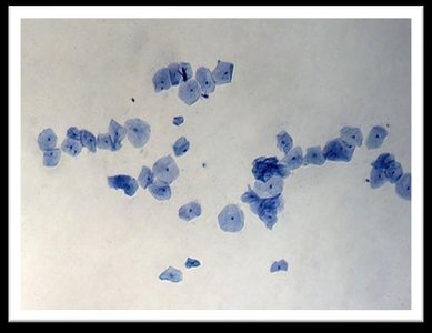
Squamous Epithelial Cells: Flat, irregularly shaped cells found in the superficial layers of the oral mucosa. They are often observed as isolated cells or in sheets due to their cohesive nature.
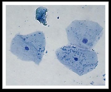
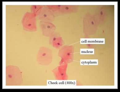
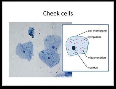
Key Features: Distinct plasma membrane, finely granular cytoplasm, and centrally or eccentrically placed nuclei.
Laboratory Preparation: Observing Cheek Cells
Cheek cells provide an accessible and illustrative example of animal cell structure. Staining with methylene blue enhances the visibility of cellular components under the microscope.
Materials: Glass slide, coverslip, toothpick, methylene blue stain, and a sample from the inner cheek.
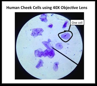
Procedure: Cells are gently scraped, mixed with stain, covered, and observed under low and then higher magnification.
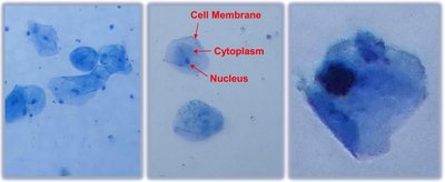
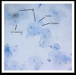
Observations: Students should identify and label the cell membrane, nucleus, and cytoplasm. Differences in cell appearance and arrangement may be noted.
Microscopic Images of Cheek Cells
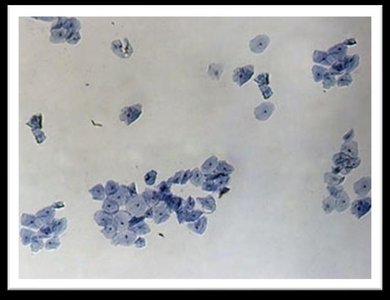
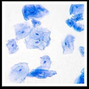
Stained cheek cells reveal characteristic features of squamous epithelial cells, including their polygonal shape, prominent nuclei, and granular cytoplasm.
Summary Table: Key Features of Squamous Epithelial Cells
Feature | Description |
|---|---|
Shape | Irregular polygon, flat |
Membrane | Distinct plasma membrane |
Cytoplasm | Finely granular, stains blue with methylene blue |
Nucleus | Small, round or oval, centrally or eccentrically placed |
Arrangement | Isolated or in cohesive sheets |
Additional info:
Cheek cells are a classic example of animal cells used in introductory cell biology labs due to their accessibility and clear demonstration of basic cell structure.
Proper staining and focusing techniques are essential for visualizing cellular details.
