 Indietro
The Cell: Structure, Function, and Origins – A Comprehensive Overview
Guida di studio - Note intelligenti
 Appunti personalizzati basati sui tuoi materiali, ampliati con definizioni chiave, esempi e contesto.
4.1 The Origins of the First Cells
Abiotic Synthesis of Organic Molecules
The origin of life on Earth is hypothesized to have occurred through a series of chemical and physical processes that led to the formation of the first cells. Scientists propose four main phases: (1) abiotic synthesis of simple organic compounds, (2) polymerization of these monomers into macromolecules, (3) emergence of a self-replicating informational molecule, and (4) encapsulation within a membrane to form a primitive cell.
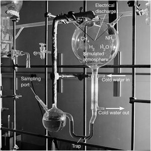
Abiotic Synthesis: Laboratory experiments, such as the Miller-Urey experiment, demonstrated that simple organic molecules like amino acids can form under simulated early Earth conditions using energy sources such as lightning or UV radiation.
Key Molecules: Amino acids, nucleic acid bases, and simple sugars are among the compounds that can be synthesized abiotically.
Energy Sources: Lightning, UV radiation, and hydrothermal vents are considered possible energy sources for these reactions.
Experimental Evidence: The Miller apparatus showed that exposing a mixture of gases (CH4, NH3, H2O, H2) to electrical discharge can yield amino acids and other organic compounds.
Additional info: Modern analyses have revealed that a wider variety of organic molecules can be produced under these conditions, supporting the plausibility of abiotic synthesis on early Earth.
The RNA World Hypothesis
RNA is hypothesized to have been the first informational molecule due to its ability to both store genetic information and catalyze chemical reactions (ribozymes).
Ribozymes: Catalytic RNAs capable of self-replication and other enzymatic activities.
Support: Modern cells use ribosomal RNA to catalyze peptide bond formation, suggesting an ancient role for RNA.
Limitations: Laboratory ribozymes have not yet achieved full self-replication, and the origin of the genetic code remains unresolved.
Formation of Protocells
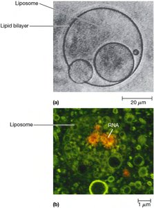
The encapsulation of self-replicating molecules within lipid membranes likely led to the first protocells. Liposomes, artificial vesicles formed from lipids, can spontaneously assemble and encapsulate macromolecules, providing a model for primitive cell membranes.
Liposomes: Hollow, membrane-bound vesicles that can grow, divide, and encapsulate RNA and other molecules.
Role of Minerals: Clays like montmorillonite may have catalyzed vesicle formation and concentrated organic molecules.
4.2 Basic Properties of Cells
The Three Domains of Life
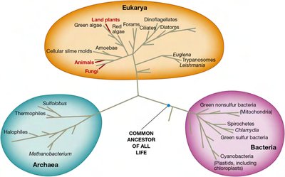
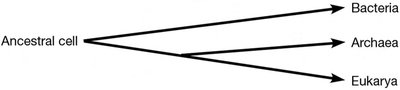
All living organisms are classified into three domains based on molecular and genetic evidence: Bacteria, Archaea, and Eukarya. This classification reflects fundamental differences in cellular structure, biochemistry, and genetics.
Bacteria: Single-celled, non-nucleated organisms with peptidoglycan cell walls.
Archaea: Single-celled, non-nucleated organisms with unique membrane lipids and often found in extreme environments.
Eukarya: Organisms with membrane-bounded nuclei and complex organelles, including plants, animals, fungi, and protists.
Comparative Properties of Bacteria, Archaea, and Eukaryotes
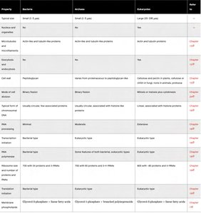
The three domains share some features but also have distinct differences in cell structure, genetic organization, and metabolism.
Property | Bacteria | Archaea | Eukaryotes |
|---|---|---|---|
Typical size | Small (1–5 μm) | Small (1–5 μm) | Large (10–100 μm) |
Nucleus and organelles | No | No | Yes |
Internal membranes | Rare | Rare | Extensive |
Cell wall composition | Peptidoglycan | Varied (no peptidoglycan) | Cellulose (plants), chitin (fungi), or none (animals) |
DNA organization | Circular, few proteins | Circular, histone-like proteins | Linear, histones |
Ribosome size | 70S | 70S | 80S |
Reproduction | Binary fission | Binary fission | Mitosis/meiosis |
Cell Size Limitations
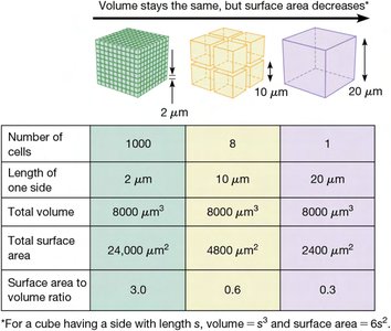
Cell size is constrained by the surface area-to-volume ratio, diffusion rates, and the need to maintain adequate concentrations of reactants and enzymes.
Surface Area/Volume Ratio: As cells grow, volume increases faster than surface area, limiting exchange with the environment.
Diffusion Rates: Larger cells face slower diffusion of molecules, which can limit metabolic efficiency.
Compartmentalization: Eukaryotic cells use organelles to localize and concentrate biochemical reactions.
Prokaryotic and Eukaryotic Cell Structure
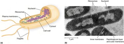
Bacteria and archaea lack membrane-bounded organelles and nuclei, while eukaryotes possess complex internal compartmentalization.
Nucleoid vs. Nucleus: Prokaryotes have a nucleoid region; eukaryotes have a true nucleus.
Internal Membranes: Eukaryotes have extensive internal membranes (e.g., ER, Golgi, mitochondria).
Membrane Trafficking and Vesicle Transport
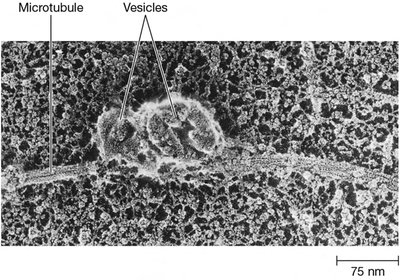
Eukaryotic cells use endocytosis and exocytosis for material exchange and vesicle transport for intracellular trafficking.
Genetic Organization and Expression
Genetic material is organized and expressed differently in the three domains:
Bacteria: Single circular chromosome, minimal RNA processing, polycistronic mRNA.
Archaea: Circular DNA, moderate RNA processing, similarities to eukaryotic transcription/translation.
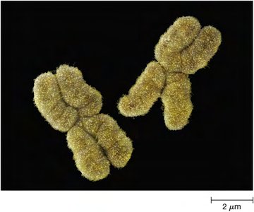
Eukaryotes: Multiple linear chromosomes, extensive RNA processing, monocistronic mRNA.
4.3 The Eukaryotic Cell in Overview: Structure and Function
Major Structural Features of Eukaryotic Cells
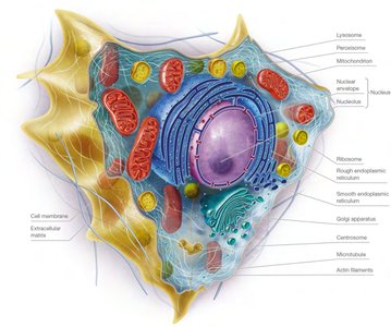
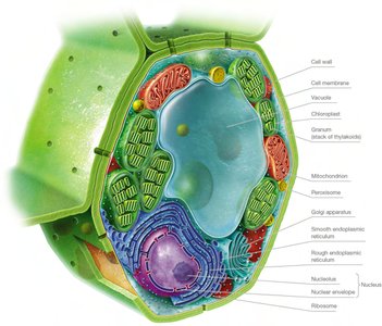
Eukaryotic cells are characterized by a plasma membrane, nucleus, membrane-bounded organelles, and a cytoskeleton. Plant and fungal cells also have a rigid cell wall, while animal cells are surrounded by an extracellular matrix.
The Plasma Membrane
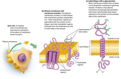
The plasma membrane defines cell boundaries and retains cellular contents. It is composed of a phospholipid bilayer with embedded proteins, many of which are glycoproteins involved in transport, signaling, and structural support.
The Nucleus
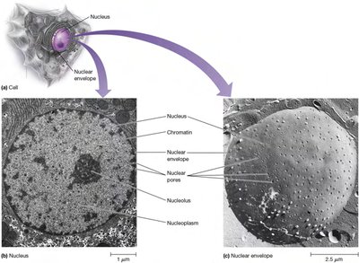
The nucleus houses the cell's genetic material and is surrounded by a double membrane called the nuclear envelope, which contains nuclear pores for regulated exchange of materials. The nucleolus within the nucleus is responsible for ribosomal RNA synthesis and ribosome assembly.
Mitochondria and Chloroplasts
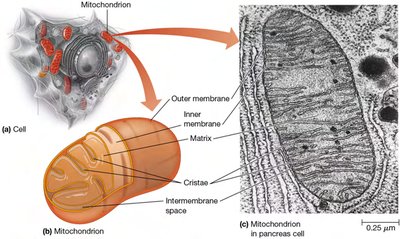
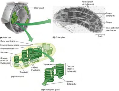
Mitochondria are the sites of aerobic respiration and ATP production, while chloroplasts carry out photosynthesis in plants and algae. Both organelles contain their own DNA and ribosomes, supporting the endosymbiont theory of their origin from ancestral bacteria.
Endosymbiont Theory and Eukaryotic Origins
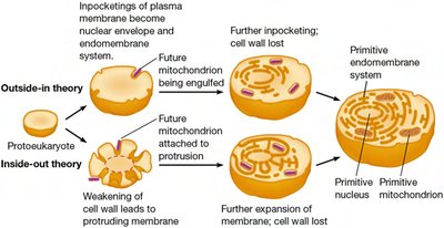
The endosymbiont theory proposes that mitochondria and chloroplasts originated from free-living bacteria that were engulfed by ancestral eukaryotic cells. Competing models (outside-in vs. inside-out) describe different mechanisms for the development of internal membranes and organelles.
The Endomembrane System
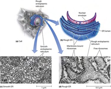
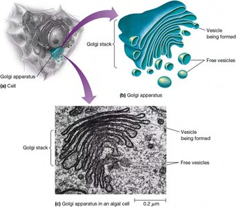
The endomembrane system includes the endoplasmic reticulum (ER), Golgi apparatus, lysosomes, and vesicles. It is responsible for the synthesis, processing, and transport of proteins and lipids.
Rough ER: Studded with ribosomes; site of membrane and secretory protein synthesis.
Smooth ER: Involved in lipid synthesis and detoxification.
Golgi Apparatus: Modifies, sorts, and packages proteins and lipids for delivery.
Lysosomes: Contain hydrolytic enzymes for intracellular digestion.
Other Organelles: Peroxisomes and Vacuoles
Peroxisomes are involved in the breakdown of fatty acids and detoxification of harmful substances. Vacuoles serve as storage and help maintain turgor pressure in plant cells.
Ribosomes and Protein Synthesis
Ribosomes are the sites of protein synthesis and are found in all cells. Eukaryotic ribosomes are larger (80S) than prokaryotic ribosomes (70S).
The Cytoskeleton
The cytoskeleton is a dynamic network of protein filaments (microtubules, microfilaments, intermediate filaments) that provides structural support, facilitates cell movement, and organizes intracellular transport.
Extracellular Structures
Animal cells are supported by an extracellular matrix (ECM) composed of proteins and polysaccharides. Plant and fungal cells have rigid cell walls made primarily of cellulose or chitin, respectively. Bacterial cell walls are composed of peptidoglycan.
4.4 Viruses, Viroids, and Prions: Agents That Invade Cells
Viruses
Viruses are acellular infectious agents consisting of a nucleic acid core (DNA or RNA) surrounded by a protein coat (capsid). They lack metabolism and reproduce only by hijacking host cell machinery.
Structure: Simple viruses have a single nucleic acid and protein type; complex viruses may have multiple types and an envelope derived from host membranes.
Examples: Influenza virus, HIV, bacteriophages.
Viroids
Viroids are small, circular RNA molecules that infect plants. They do not encode proteins but can replicate within host cells and cause disease by interfering with gene expression.
Prions
Prions are infectious proteins that cause neurodegenerative diseases by inducing abnormal folding of normal cellular proteins. Examples include scrapie in sheep, mad cow disease in cattle, and Creutzfeldt-Jakob disease in humans.
These study notes provide a comprehensive overview of cell structure, function, and origins, integrating experimental evidence, comparative cell biology, and the roles of acellular infectious agents. They are designed to support exam preparation and foundational understanding for college-level Cell Biology.
