 Indietro
The Endomembrane System and Protein Sorting: ER and Golgi Roles in Protein Processing and Trafficking
Guida di studio - Note intelligenti
 Appunti personalizzati basati sui tuoi materiali, ampliati con definizioni chiave, esempi e contesto.
The Endomembrane System and Protein Sorting
Roles of the ER and Golgi in Protein Processing
The endoplasmic reticulum (ER) and Golgi apparatus are central to the processing and modification of proteins within eukaryotic cells. These organelles facilitate protein folding, quality control, and glycosylation, which is the addition of carbohydrate side chains to proteins, forming glycoproteins.
Protein Folding: Newly synthesized polypeptides undergo folding in the ER, assisted by chaperone proteins such as calnexin and calreticulin.
Quality Control: Misfolded proteins are identified and retained in the ER for refolding or degradation.
Glycosylation: The addition of oligosaccharide chains to proteins, which occurs in two main forms:
N-linked glycosylation: Attachment of oligosaccharides to the nitrogen atom of asparagine residues.
O-linked glycosylation: Attachment of oligosaccharides to the oxygen atom of serine, threonine, or rarely tyrosine residues.
Core Oligosaccharide: All carbohydrate side chains initially have a common core consisting of two N-acetylglucosamine, nine mannose, and three glucose units.
Example: Glycoproteins such as antibodies and cell surface receptors rely on proper glycosylation for their function.
Initial Glycosylation in the ER
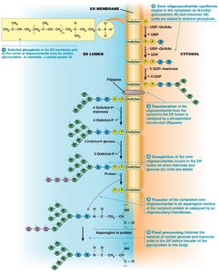
The first steps of N-linked glycosylation occur on the cytosolic side of the ER membrane, followed by further processing in the ER lumen. Dolichol phosphate acts as an oligosaccharide carrier, facilitating the assembly and transfer of the core oligosaccharide to the recipient protein.
Dolichol Phosphate: Embedded in the ER membrane, it receives N-acetylglucosamine and mannose groups.
Flippase: Translocates the growing oligosaccharide to the ER lumen.
Transfer to Protein: The completed oligosaccharide is transferred to an asparagine residue of the protein.
Trimming and Modification: The attached oligosaccharide is further processed.
Cotranslational Glycosylation and Protein Folding
Most glycosylation occurs cotranslationally, meaning the oligosaccharide is added as the polypeptide is being synthesized. This process promotes proper folding, assisted by calnexin and calreticulin, which bind monoglucosylated glycoproteins and facilitate disulfide bond formation.
UGGT: A glucosyl transferase that adds glucose to improperly folded proteins, allowing further folding attempts.
Glucosidase II: Removes the final glucose after proper folding is achieved.
Example: Defects in ER glycosylation can lead to diseases such as congenital disorders of glycosylation.
Further Glycosylation in the Golgi Complex
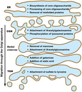
As glycoproteins move from the cis to the trans face of the Golgi, terminal glycosylation occurs, creating diversity in glycoprotein structures. Hundreds of glycosyl transferases in the ER and Golgi attach various carbohydrate groups to proteins.
Terminal Glycosylation: Removal or addition of monosaccharides such as N-acetylglucosamine, galactose, or sialic acid.
Enzyme Diversity: The Golgi contains many glycosyl transferases, each with specific substrate preferences.
Protein Trafficking: ER and Golgi Roles
Proteins synthesized in the rough ER are directed to various cellular locations by specific tags, which may be amino acid sequences, hydrophobic domains, or oligosaccharide side chains. These tags ensure proper sorting and retention within the endomembrane system.
Protein Tags: Direct proteins to their correct destinations and prevent their escape.
Overview: Nuclear genes are transcribed, processed, and translated, after which polypeptides are sorted via two main pathways.
Pathways for Sorting Tagged Polypeptides
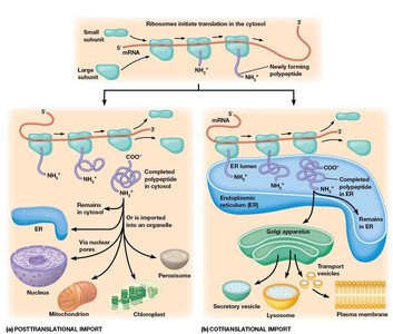
There are two main pathways for sorting newly synthesized polypeptides:
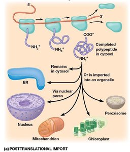
Posttranslational Import: Polypeptides destined for the cytosol, mitochondria, chloroplast, peroxisome, or nucleus are synthesized on free ribosomes and imported after translation.
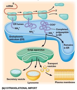
Cotranslational Import: Polypeptides destined for the endomembrane system or export are synthesized on ribosomes attached to the ER membrane and imported during translation.
Trafficking Through the Endomembrane System
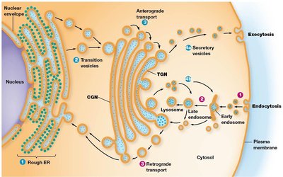
Proteins and lipids move through the endomembrane system via vesicular transport. Anterograde transport moves materials from the ER to the Golgi and then to the plasma membrane or lysosomes, while retrograde transport returns compartment-specific proteins to earlier compartments.
Anterograde Transport: Transition vesicles carry proteins from the ER to the Golgi; secretory vesicles release contents by exocytosis.
Retrograde Transport: Endocytosis brings materials into the cell; retrograde traffic returns proteins to the ER or Golgi.
Cotranslational Import and the Role of the ER in Protein Synthesis
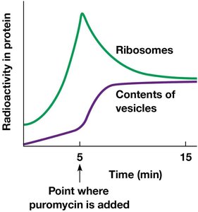
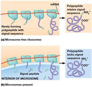
Cotranslational import is essential for delivering proteins to the endomembrane system. Experiments with microsomes demonstrated that newly formed polypeptides pass into the ER lumen during synthesis, allowing proper routing and processing.
Microsomes: Vesicle-like artifacts formed from rough ER fragments used to study protein synthesis.
Puromycin: An antibiotic used to halt protein synthesis and analyze polypeptide localization.
Signal Hypothesis and ER Signaling Sequences
The signal hypothesis states that polypeptides destined for the ER contain an N-terminal signal sequence that directs the ribosome-mRNA-polypeptide complex to the ER surface. The sequence is typically 15–30 amino acids long, with a positively charged N-terminal region, a central hydrophobic region, and a polar region near the cleavage site.
Preproteins: Proteins containing ER signal sequences, such as preproinsulin.
Translocon: A structure in the ER membrane responsible for translocation of polypeptides.
SRP Mechanism of Cotranslational Import
The signal recognition particle (SRP) recognizes and binds to the ER signal sequence, halting translation and directing the ribosome to the ER membrane. The ribosome docks at the translocon, and the elongating peptide passes into the ER lumen, where the signal sequence is cleaved.
SRP: Binds the ER signal sequence and ribosome, mediating attachment to the ER membrane.
Signal Peptidase: Cleaves the ER signal sequence after translocation.
Protein Routing After ER Entry
Proteins released into the ER lumen are routed to the Golgi apparatus, secretory vesicles, or back to the ER. Soluble proteins may be secreted or targeted to other parts of the endomembrane system by specific carbohydrate side chains or amino acid sequences.
Retention Tags: Sequences such as RXR prevent proteins from leaving the ER.
Retrieval Tags: Short C-terminal sequences (e.g., KDEL, KKXX) ensure proteins are returned from the Golgi to the ER.
Example: The KDEL sequence is critical for the retrieval of ER-resident proteins.
Golgi Complex Protein Sorting
Golgi-specific proteins are integral membrane proteins with one or more membrane-spanning domains. The length of the hydrophobic domains determines their incorporation into specific Golgi cisternae, as membrane thickness increases from the ER to the plasma membrane.
Membrane Thickness: Increases from 5 nm (ER) to 8 nm (plasma membrane).
Hydrophobic Domain Length: Correlates with protein localization within the Golgi.
Targeting of Lysosomal Proteins
Soluble lysosomal enzymes undergo N-glycosylation in the ER and Golgi, followed by phosphorylation of mannose residues to form mannose-6-phosphate. This tag ensures delivery of lysosomal proteins to lysosomes.
Mannose-6-Phosphate: A targeting signal for lysosomal enzymes.
Insertion of Integral Membrane Proteins
Integral membrane proteins are anchored to the ER membrane by stop-transfer sequences or signal sequences. Once incorporated, they may remain in the ER or be transported to other endomembrane compartments.
Stop-Transfer Sequences: Mediate insertion and anchoring of membrane proteins.
Transport: Occurs via membrane budding and fusion events.
Posttranslational Import into the ER Lumen
Some proteins are synthesized in the cytosol and imported into the ER lumen posttranslationally. Chaperones such as Hsp70 keep the protein unfolded, and a complex of ER membrane proteins (Sec62, 63, 71, 72) targets the protein for translocation. BiP couples ATP hydrolysis to pull the polypeptide into the ER lumen.
Chaperones: Prevent premature folding during import.
Sec Complex: Facilitates translocation across the ER membrane.
BiP: Provides energy for import via ATP hydrolysis.
Example: Posttranslational import is essential for the proper localization of certain secretory and membrane proteins.
Summary Table: Glycosylation and Protein Sorting Steps
Location | Key Processes |
|---|---|
ER | Biosynthesis and processing of core oligosaccharide, removal of misfolded proteins |
CGN (cis Golgi network) | Attachment of N-acetylgalactosamine, phosphorylation of lysosomal proteins |
Medial cisternae | Removal of mannose, attachment of N-acetylglucosamine |
TGN (trans Golgi network) | Attachment of sulfate to tyrosine |
Additional info: The notes have been expanded to include definitions, examples, and context for each step in protein processing and trafficking, as well as a summary table for glycosylation steps.
