 Indietro
The Endomembrane System and Protein Sorting: Exocytosis, Endocytosis, and Vesicular Transport
Guida di studio - Note intelligenti
 Appunti personalizzati basati sui tuoi materiali, ampliati con definizioni chiave, esempi e contesto.
The Endomembrane System and Protein Sorting
Overview of Exocytosis and Endocytosis
The endomembrane system is a network of membranes within eukaryotic cells that facilitates the transport, sorting, and secretion of proteins and lipids. Two key processes, exocytosis and endocytosis, are unique to eukaryotes and enable the movement of materials across the plasma membrane.
Exocytosis: The process by which secretory vesicles release their contents outside the cell.
Endocytosis: The process by which cells internalize external materials by forming vesicles from the plasma membrane.
Secretory Pathways: Transport to the Cell Exterior
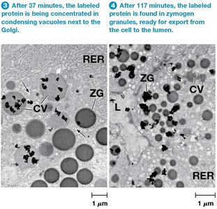
Proteins synthesized in the rough endoplasmic reticulum (ER) are transported through the Golgi complex to secretory vesicles and granules, which discharge their contents outside the cell. Experimental evidence from electron microscopy and autoradiography demonstrates the sequential movement of proteins through these pathways.
Step 1: Proteins are radioactively labeled and initially detected in the rough ER.
Step 2: Labeled proteins appear in the Golgi apparatus after a few minutes.
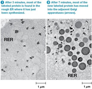
Step 3: After 37 minutes, proteins are found in vesicles budding from the Golgi (condensing vacuoles).
Step 4: After 117 minutes, proteins accumulate in dense zymogen granules, ready for secretion.
Constitutive and Regulated Secretion
Secretory vesicles follow two main pathways:
Constitutive Secretion: Vesicles move directly to the cell surface and fuse with the plasma membrane continuously, independent of external signals. Example: mucus secretion by intestinal lining.
Regulated Secretion: Vesicles accumulate in the cell and fuse with the plasma membrane only in response to specific signals (e.g., neurotransmitter or insulin release).
Polarized Secretion
Exocytosis can be restricted to specific surfaces of polarized cells. For example, intestinal cells secrete digestive enzymes only on the side facing the intestine.
The Process of Exocytosis
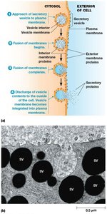
Exocytosis involves the movement of vesicles to the cell surface, fusion with the plasma membrane, and release of contents. The vesicle membrane becomes part of the plasma membrane.
Step 1: Vesicle approaches the plasma membrane.
Step 2: Fusion of membranes occurs.
Step 3: Discharge of vesicle contents to the exterior.
Step 4: Vesicle membrane is integrated into the plasma membrane.
Orientation of Membrane Components
Upon fusion, the lumenal (inner) membrane of the vesicle becomes part of the outer surface of the plasma membrane. Glycolipids and glycoproteins formed in the ER and Golgi lumens face the extracellular space.
Mechanism and Regulation of Exocytosis
Microtubules: Vesicle movement is dependent on microtubules; colchicine inhibits this process.
Calcium: A transient elevation in Ca2+ is essential for triggering regulated exocytosis.
Endocytosis: Importing Extracellular Molecules
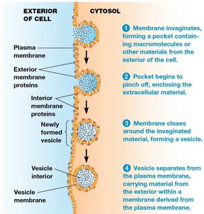
Endocytosis allows cells to internalize extracellular material by forming vesicles from the plasma membrane. This process is essential for nutrient uptake, receptor recycling, and defense against pathogens.
Step 1: Plasma membrane invaginates, forming a pocket.
Step 2: Pocket pinches off, enclosing material.
Step 3: Membrane closes around the material, forming a vesicle.
Step 4: Vesicle separates from the plasma membrane.
Types and Functions of Endocytosis
Phagocytosis: Ingestion of large particles or microorganisms by specialized cells (phagocytes).
Pinocytosis: Uptake of liquids.
Receptor-Mediated Endocytosis: Specific uptake of macromolecules via cell surface receptors.
Endocytosis is involved in maintaining cell homeostasis, nutrient uptake, receptor recycling, protection from pathogens, and intercellular communication.
Phagocytosis and Immune Function
Phagocytosis is a key defense mechanism in multicellular organisms. Neutrophils and macrophages engulf and digest foreign materials, cellular debris, and damaged cells.
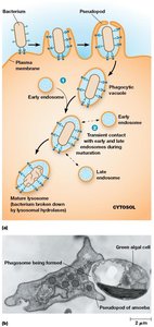
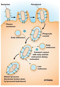
Phagocytosis in Amoebae
Amoebae use phagocytosis for nutrition. Contact with food triggers pseudopod formation, engulfing the object and forming a phagosome, which fuses with lysosomes for digestion.
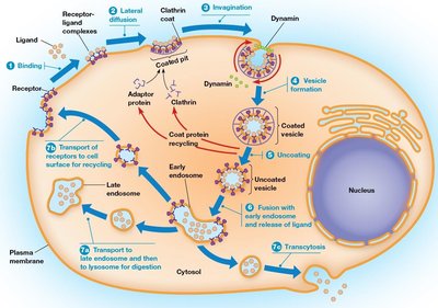
Receptor-Mediated Endocytosis
This process allows cells to internalize specific substances by binding them to cell surface receptors, concentrating them in coated pits, and forming coated vesicles.
Step 1: Ligands bind to receptors.
Step 2: Receptor-ligand complexes accumulate in coated pits.
Step 3: Adaptor proteins, clathrin, and dynamin induce curvature and invagination.
Step 4: Pit pinches off, forming a coated vesicle.
Step 5: Clathrin coat is released, leaving an uncoated vesicle.
Step 6: Vesicle fuses with an early endosome.
Step 7: Material is transported to late endosome/lysosome, receptors are recycled, or material is transcytosed.
Clathrin-Independent Endocytosis
Fluid-phase endocytosis is a nonspecific process for internalizing extracellular fluid, compensating for membrane segments added by exocytosis.
Coated Vesicles in Cellular Transport
Most vesicles involved in protein and lipid transport are coated with proteins such as clathrin, COPI, and COPII. The type of coat protein determines vesicle destination and induces curvature for vesicle formation.
Clathrin: Forms lattices around vesicles, driving their formation.
COPI: Involved in retrograde transport from Golgi to ER.
COPII: Mediates transport from ER to Golgi.
Structure and Function of Clathrin-Coated Vesicles
Clathrin-coated vesicles are surrounded by lattices composed of clathrin and adaptor proteins. The basic unit is the triskelion, a three-legged structure that assembles into hexagons and pentagons, allowing the coat to curve around budding vesicles.
Adaptor Protein Complexes
Adaptor protein (AP) complexes promote assembly of clathrin coats and regulate clathrin assembly/disassembly. There are four types of AP complexes, each binding different receptors.
Clathrin Coat Assembly and Vesicle Formation
Clathrin units initially form a planar hexagonal structure. As more triskelions are incorporated, pentagons form, allowing the coat to curve and bud off as a vesicle. Dynamin, a GTPase, is required for constricting and closing the vesicle.
Uncoating of Vesicles
Clathrin dissociates rapidly after vesicle formation, a process requiring energy from an uncoating ATPase.
COPI- and COPII-Coated Vesicles: ER-Golgi Transport
COPI-coated vesicles mediate retrograde transport from Golgi to ER and bidirectional transport between Golgi cisternae. COPII-coated vesicles mediate anterograde transport from ER to Golgi. Proper sorting and targeting involve SNARE proteins:
v-SNAREs: Found on vesicles.
t-SNAREs: Found on target membranes.
Complementary v- and t-SNAREs allow recognition and fusion between vesicles and targets.
Summary Table: Types of Vesicle Coats and Their Functions
Coat Protein | Main Function | Destination |
|---|---|---|
Clathrin | Endocytosis, vesicle formation from plasma membrane and TGN | Endosomes, lysosomes |
COPI | Retrograde transport | Golgi to ER, intra-Golgi |
COPII | Anterograde transport | ER to Golgi |
Caveolin | Formation of caveolae, cholesterol uptake | Various, including plasma membrane |
Additional info: The SNARE hypothesis explains the specificity of vesicle targeting and fusion, which is essential for maintaining compartmentalization and proper protein sorting in the cell.
