 Indietro
Amino Acids and Proteins: Structure, Properties, and Spectroscopy
Guida di studio - Note intelligenti
 Appunti personalizzati basati sui tuoi materiali, ampliati con definizioni chiave, esempi e contesto.
Amino Acids: The Building Blocks of Proteins
Structure of Amino Acids
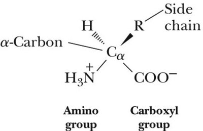
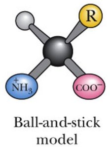
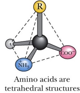
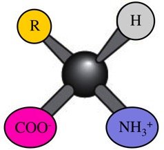
Amino acids are organic molecules that serve as the fundamental units of proteins. Each amino acid consists of a central α-carbon (Cα) bonded to four distinct groups: an amino group (NH3+), a carboxyl group (COO-), a hydrogen atom, and a unique side chain (R group) that determines the amino acid's properties.
α-Carbon: The central carbon atom to which all groups are attached.
Amino group: Acts as a base, accepting protons.
Carboxyl group: Acts as an acid, donating protons.
Side chain (R group): Varies among amino acids, conferring specific chemical characteristics.
Polymerization of Amino Acids
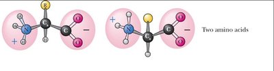
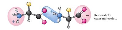
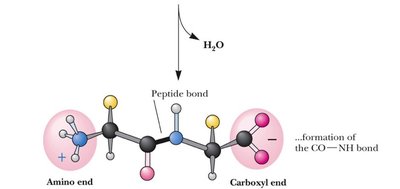
Amino acids link together via peptide bonds to form polypeptides and proteins. The process involves the removal of a water molecule (condensation reaction) and results in a chain with an N-terminus (amino end) and a C-terminus (carboxyl end).
Peptide bond: Covalent bond formed between the carboxyl group of one amino acid and the amino group of another.
N-terminus: The end of the polypeptide with a free amino group.
C-terminus: The end of the polypeptide with a free carboxyl group.
Classification of Amino Acids
Types of Amino Acids Based on Side Chains
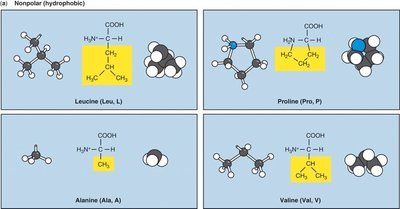
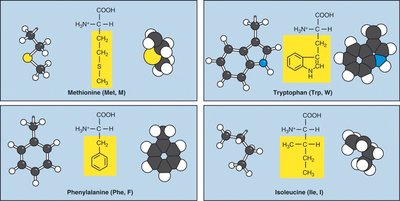
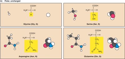
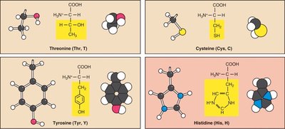
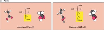
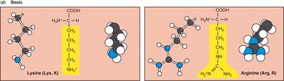
The 20 standard amino acids are classified according to the chemical nature of their side chains:
Non-polar (hydrophobic): Side chains are mostly hydrocarbons, repelling water.
Polar (hydrophilic): Side chains contain electronegative atoms, attracting water.
Acidic: Side chains have carboxyl groups, negatively charged at physiological pH.
Basic: Side chains have amino groups, positively charged at physiological pH.
Each amino acid is known by its full name, three-letter abbreviation, and one-letter code (e.g., Glycine: Gly, G).
Ionization and pKa Values
Ionizable Groups and pKa
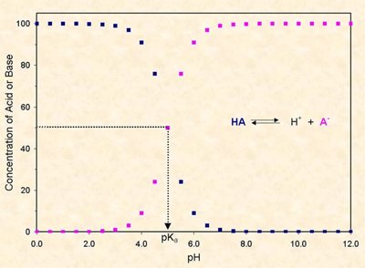
Amino acids contain ionizable groups that can gain or lose protons depending on the pH of the environment. The pKa is the pH at which half of the molecules are ionized. The charge of an amino acid changes as the pH crosses its pKa value.
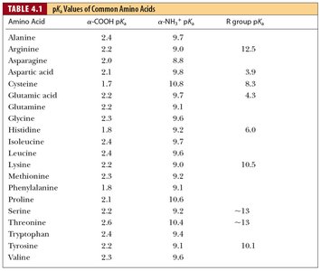
Carboxyl group: pKa ~2; ionized (COO-) at physiological pH.
Amino group: pKa ~9.5; protonated (NH3+) at physiological pH.
Side chains: pKa varies, affecting the overall charge of the amino acid.
Charge States at Physiological pH
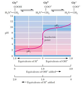
At pH 7, the backbone carboxyl group is deprotonated (negative), and the amino group is protonated (positive), resulting in a net neutral charge for the backbone. The side chain's ionization determines the overall charge of the amino acid.
Example: Glycine has a net charge of 0 at pH 7.
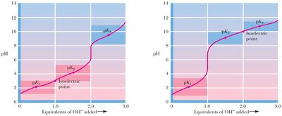
Example: Glutamic acid has a net charge of -1 at pH 7 due to its side chain.
Example: Lysine has a net charge of +1 at pH 7 due to its side chain.

Isoelectric Point and Protein Charge
Isoelectric Point (pI)
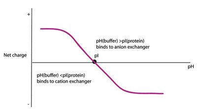
The isoelectric point (pI) is the pH at which a protein or amino acid has no net charge. The pI is important for techniques such as ion exchange chromatography, where proteins are separated based on their charge at different pH values.
Anion exchange chromatography: Proteins bind to positively charged columns if pH > pI.
Cation exchange chromatography: Proteins bind to negatively charged columns if pH < pI.
Chirality and Stereoisomers
Chiral Centers in Amino Acids
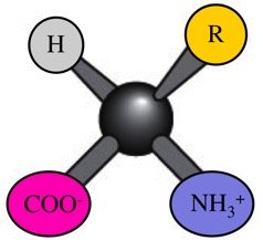
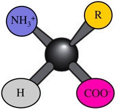
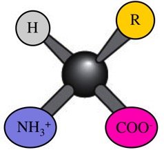
The α-carbon of most amino acids is a chiral center, meaning it is attached to four different groups. This gives rise to two stereoisomers: L and D forms. Only L-amino acids are found in proteins synthesized by living organisms.
Chirality: Property of a molecule that cannot be superimposed on its mirror image.
CORN Law: Used to distinguish L and D isomers based on the arrangement of CO, R, and N groups.
Glycine: Not chiral, as it has two identical groups attached to the α-carbon.
Isoleucine and threonine: Have two chiral centers.


Spectroscopy of Amino Acids
Optical Activity and Polarization
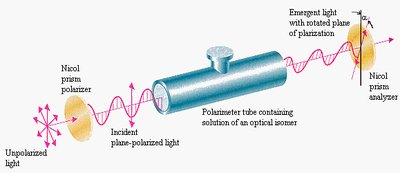
Chiral amino acids are optically active, meaning they can rotate the plane of polarized light. This property is used to distinguish between stereoisomers and is fundamental in understanding molecular interactions in biology.
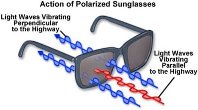
Polarization: Light oscillates in a specific plane; polarizers can filter light based on its polarization.
Optical activity: Rotation of polarized light by chiral molecules.
Racemic mixture: Equal amounts of L and D isomers, produced in non-biological synthesis.
Absorbance and Fluorescence Spectroscopy
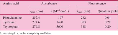
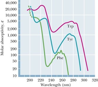
Amino acids and proteins absorb ultraviolet (UV) light, especially those with aromatic side chains (phenylalanine, tyrosine, tryptophan). Fluorescence occurs when certain amino acids re-emit absorbed light at longer wavelengths.
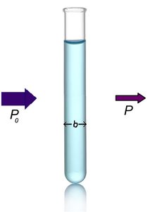
Absorbance: Measured using the Beer-Lambert Law:
Fluorescence: Emission of light by molecules after absorbing UV light; used to study protein folding and ligand binding.
Quantum yield: Efficiency of fluorescence emission.
Summary Table: pKa Values of Common Amino Acids
The following table summarizes the pKa values for the α-COOH, α-NH3+, and R group of common amino acids:
Amino Acid | α-COOH pKa | α-NH3+ pKa | R group pKa |
|---|---|---|---|
Alanine | 2.4 | 9.7 | |
Arginine | 2.2 | 9.0 | 12.5 |
Asparagine | 2.0 | 8.8 | |
Aspartic acid | 2.1 | 9.8 | 3.9 |
Cysteine | 1.7 | 10.8 | 8.3 |
Glutamic acid | 2.2 | 9.7 | 4.3 |
Glutamine | 2.2 | 9.0 | |
Glycine | 2.3 | 9.6 | |
Histidine | 1.8 | 9.3 | 6.0 |
Isoleucine | 2.2 | 9.7 | 10.5 |
Leucine | 2.4 | 9.6 | |
Lysine | 2.2 | 9.0 | 10.5 |
Methionine | 2.3 | 9.2 | |
Phenylalanine | 1.8 | 9.1 | |
Proline | 2.0 | 10.6 | |
Serine | 2.2 | 9.2 | ~13 |
Threonine | 2.1 | 9.1 | ~13 |
Tryptophan | 2.4 | 9.4 | |
Tyrosine | 2.2 | 9.1 | 10.1 |
Valine | 2.3 | 9.6 |
Key Equations
Henderson-Hasselbalch Equation:
Beer-Lambert Law:
Conclusion
Amino acids are central to the structure and function of proteins. Their chemical properties, ionization states, and optical activity are fundamental to understanding protein biochemistry and genetics. Spectroscopic techniques provide powerful tools for studying protein structure, folding, and interactions.
