 Indietro
Chapter 9: The Molecular Biology of Translation – Genetics Study Notes
Guida di studio - Note intelligenti
 Appunti personalizzati basati sui tuoi materiali, ampliati con definizioni chiave, esempi e contesto.
Polypeptides and Amino Acids
Polypeptide Composition and Assembly
Polypeptides are linear chains of amino acids assembled at ribosomes, serving as the primary structure of proteins. The diversity of amino acids allows for a wide range of chemical properties and biological functions.
Twenty amino acids are the building blocks of polypeptides.
Covalent peptide bonds form between the carboxyl group of one amino acid and the amino group of another.
Distinctive R-groups confer unique chemical properties, influencing hydrophobicity, polarity, and reactivity.
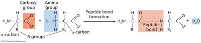
Amino Acid Structure
Each amino acid contains a central carbon (α-carbon), an amino group, a carboxyl group, and a unique R-group. The ribosome catalyzes peptide bond formation during translation.
R-groups determine the chemical behavior of each amino acid.
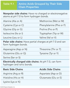
Some R-groups are nonpolar, polar, or electrically charged.
Amino Acid Classification
Amino acids are grouped based on their side chain properties, which affect protein folding and function.
Group | Examples |
|---|---|
Nonpolar side chains | Alanine, Methionine, Glycine, Phenylalanine, etc. |
Polar side chains | Asparagine, Threonine, Glutamine, Tyrosine, Serine |
Electrically charged side chains | Arginine, Aspartate, Histidine, Glutamate, Lysine |
Polypeptide and Transcript Structure
Ribosome Function and mRNA Translation
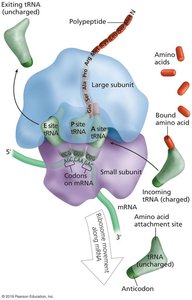
Ribosomes are complex molecular machines that translate mRNA into polypeptides by reading codons in the 5' to 3' direction.
Ribosomes contain rRNAs and proteins.
Translation occurs codon by codon, assembling amino acids in the order specified by the mRNA.
Messenger RNA (mRNA)
The sequence of mRNA determines the amino acid sequence of the resulting polypeptide. Translation boundaries are defined by start and stop codons.
5' UTR and 3' UTR are untranslated regions flanking the coding sequence.
The start codon marks the N-terminus; the stop codon marks the C-terminus.
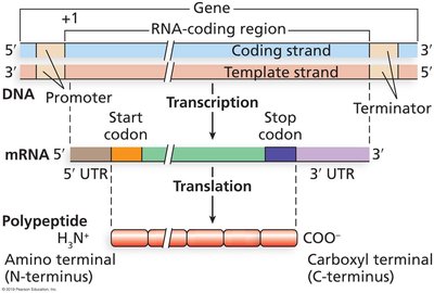
Polypeptide Structure
Levels of Polypeptide Organization
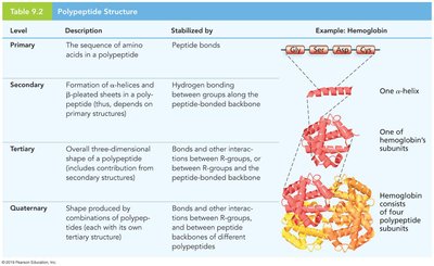
Polypeptides exhibit four levels of structural organization, each contributing to protein function.
Primary structure: Sequence of amino acids.
Secondary structure: Alpha helices and beta-pleated sheets formed by hydrogen bonding.
Tertiary structure: Three-dimensional shape due to interactions among R-groups.
Quaternary structure: Association of multiple polypeptide chains.
Level | Description | Stabilized by | Example |
|---|---|---|---|
Primary | Sequence of amino acids | Peptide bonds | Hemoglobin subunit |
Secondary | Alpha helix, beta sheet | Hydrogen bonds | One α-helix |
Tertiary | 3D shape | Bonds/interactions among R-groups | Hemoglobin subunit |
Quaternary | Multiple polypeptides | Bonds/interactions among subunits | Hemoglobin (4 subunits) |
Ribosome Structure and Function
Bacterial, Archaeal, and Eukaryotic Ribosomes
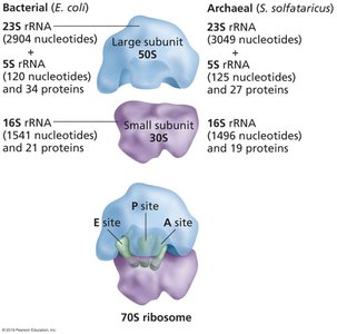
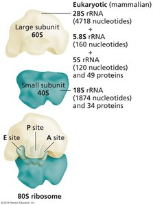
Ribosomes are composed of two subunits, each containing rRNA and proteins. Their composition and size differ among domains of life.
Bacterial ribosomes: 30S (small) + 50S (large) = 70S
Archaeal ribosomes: Similar to bacteria, but with distinct rRNA and protein composition
Eukaryotic ribosomes: 40S (small) + 60S (large) = 80S
Svedberg units (S): Measure sedimentation rate, reflecting size and shape
Ribosome Sites and Structure
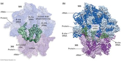
Ribosomes contain three key sites for tRNA binding: the P site (peptidyl), A site (aminoacyl), and E site (exit). The large subunit contains a channel for polypeptide emergence.
P site: Holds tRNA with attached polypeptide
A site: Binds incoming charged tRNA
E site: Exit site for uncharged tRNA
Translation Phases
Initiation
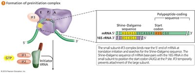
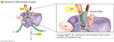
Translation initiation involves assembly of the ribosome at the start codon, guided by initiation factors and energy from GTP.
Small ribosomal subunit binds near the 5' end of mRNA.
Initiator tRNA binds to the start codon.
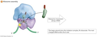
Large subunit joins, forming the intact ribosome.
Bacterial Initiation
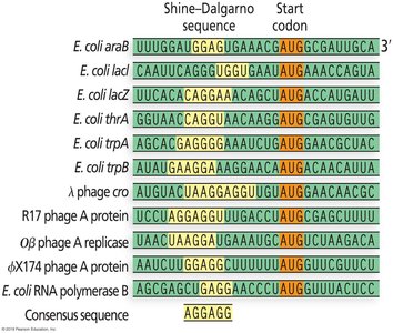
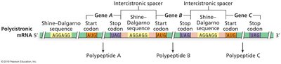
In bacteria, the Shine–Dalgarno sequence on mRNA base pairs with the 16S rRNA, guiding the ribosome to the start codon.
Six components: mRNA, small/large subunits, initiator tRNA, initiation factors, GTP.
Initiator tRNA carries N-formylmethionine (fMet).
Eukaryotic Initiation
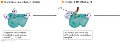
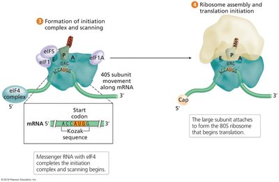
Eukaryotic initiation involves eIF proteins and scanning for the start codon, often embedded in the Kozak sequence.
Preinitiation complex forms with small subunit and eIFs.
Initiator tRNA and eIF5 join the complex.
mRNA joins, and the complex scans for the start codon (AUG).
Kozak sequence: 5'-ACCAUGG-3'
Elongation
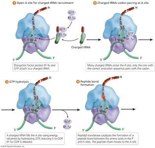
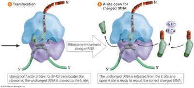
Elongation is the process of adding amino acids to the growing polypeptide chain, facilitated by elongation factors and GTP hydrolysis.
Charged tRNAs are recruited to the A site.
Peptide bonds form between amino acids at the P and A sites.
Ribosome translocates along mRNA, moving tRNAs through the sites.
Termination
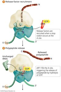
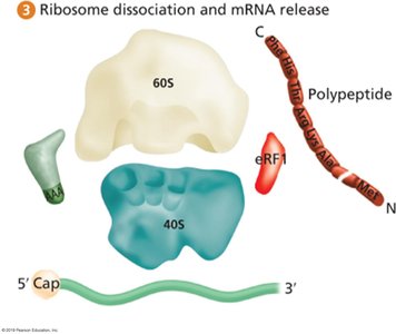
Termination occurs when a stop codon enters the A site, triggering release factors to release the polypeptide and dissociate the ribosome.
Three stop codons: UAA, UAG, UGA.
Bacterial release factors: RF1, RF2, RF3.
Eukaryotic release factors: eRF1, eRF3.
Translation Efficiency and Polyribosomes
Translational Complex and Polyribosomes
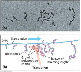
Translation is highly efficient, with multiple ribosomes (polyribosomes) translating a single mRNA simultaneously.
Bacterial cells contain ~20,000 ribosomes.
Polyribosomes increase the rate of protein synthesis.
Coupling of Transcription and Translation
In bacteria, translation can begin before transcription is complete. In eukaryotes, mRNA must be processed and exported from the nucleus.
Bacterial mRNAs are often polycistronic, encoding multiple proteins.
Eukaryotic mRNAs are typically monocistronic.
The Genetic Code
Codons and tRNA
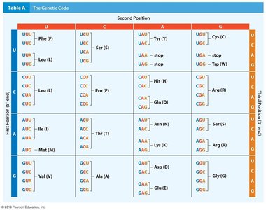
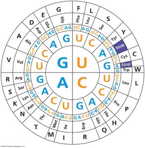
The genetic code is the correspondence between mRNA codons and amino acids. tRNAs act as adaptors, carrying amino acids and matching codons via their anticodon.
64 codons: 61 specify amino acids, 3 are stop codons.
Redundancy: Most amino acids are specified by multiple codons (synonymous codons).
Codon | Amino Acid |
|---|---|
AUG | Methionine (Start) |
UUU, UUC | Phenylalanine |
UAA, UAG, UGA | Stop |
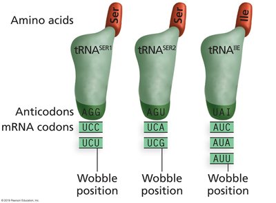
Third-Base Wobble
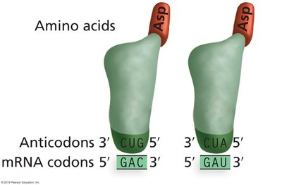
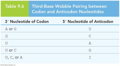
Third-base wobble allows flexibility in codon-anticodon pairing, reducing the number of tRNAs needed to decode all codons.
Isoaccepting tRNAs: Different tRNAs for the same amino acid.
Wobble occurs at the 3' base of the codon and 5' base of the anticodon.
3' Nucleotide of Codon | 5' Nucleotide of Anticodon |
|---|---|
A or G | U |
G | C |
U | A |
U or C | G |
U, C, or A | I |
Universality and Exceptions of the Genetic Code
The genetic code is nearly universal across organisms, enabling recombinant protein production. Exceptions exist, mainly in mitochondria and a few nuclear genes.
tRNA Charging and Aminoacyl-tRNA Synthetase
Charging tRNA Molecules
tRNAs are charged with their corresponding amino acids by aminoacyl-tRNA synthetases, ensuring fidelity in translation.
Each synthetase recognizes specific tRNAs (isoacceptors).
ATP provides energy for amino acid attachment.
Protein Folding and Posttranslational Processing
Posttranslational Modifications
Proteins undergo folding and chemical modifications after translation to achieve their functional forms.
Removal of N-terminal amino acids (e.g., fMet, methionine).
Phosphorylation, methylation, acetylation, and addition of carbohydrate side chains.
Cleavage into functional segments (e.g., insulin processing).
