 Indietro
Chromosome Distribution During Cell Division: Binary Fission, Mitosis, and Meiosis
Guida di studio - Note intelligenti
 Appunti personalizzati basati sui tuoi materiali, ampliati con definizioni chiave, esempi e contesto.
Chromatin, Chromosomes, and Chromosome Structure
Definitions and Key Features
Understanding the structure and function of chromosomes is fundamental to genetics. Chromatin and chromosomes are two forms of DNA organization within the cell, each with distinct characteristics and roles during the cell cycle.
Chromatin: DNA complexed with proteins (mainly histones) that exists in a less condensed form during interphase.
Chromosome: Highly condensed, compacted chromatin visible during cell division. Each chromosome consists of a single, continuous DNA molecule.
Euchromatin: Less compact chromatin, generally transcriptionally active.
Heterochromatin: Highly compact chromatin, typically transcriptionally inactive.
Parts of a Chromosome
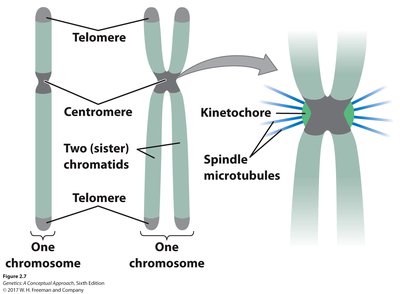
Centromere: The constricted region of a chromosome, essential for proper segregation during cell division. It is the site of kinetochore formation and spindle attachment.
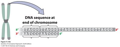
Telomeres: Repetitive DNA sequences at the ends of eukaryotic chromosomes that protect against loss of genetic information during replication. In humans, the telomeric sequence is 5'-TTAGGG-3'.
Kinetochore: Protein complex assembled at the centromere, mediating attachment to spindle microtubules during mitosis and meiosis.
DNA Replication and Chromosome Duplication
DNA Replication in the Cell Cycle
DNA replication is the process by which a cell duplicates its DNA, ensuring that each daughter cell receives an identical set of genetic material. Replication occurs during the S phase of the cell cycle.
After replication, each chromosome consists of two identical sister chromatids joined at the centromere.
The number of chromosomes remains constant, but the number of DNA molecules (chromatids) doubles until cell division occurs.
Types of Cell Division
Overview
Cells divide by several mechanisms, each with distinct outcomes and biological significance:
Binary Fission: Prokaryotic cell division.
Mitosis: Eukaryotic cell division producing genetically identical daughter cells.
Meiosis: Eukaryotic cell division producing haploid gametes, introducing genetic diversity.
Binary Fission
Mechanism and Significance
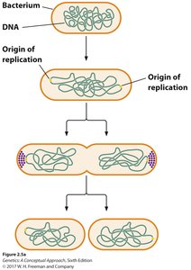
Binary fission is the primary mode of cell division in prokaryotes (bacteria and archaea), as well as in mitochondria and chloroplasts. It ensures equal distribution of genetic material to daughter cells.
DNA replication begins at a specific origin of replication.
Two DNA molecules are separated and anchored to opposite ends of the cell.
A septum forms, dividing the parent cell into two genetically identical daughter cells.
Mitosis
Purpose and Overview
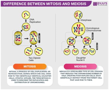
Mitosis is the process by which eukaryotic cells divide to produce two genetically identical daughter cells, maintaining the chromosome number of the parent cell. It is essential for growth, repair, and asexual reproduction.
Mitosis consists of several stages: Prophase, Prometaphase, Metaphase, Anaphase, Telophase, and Cytokinesis.
Chromatids separate, ensuring each daughter cell receives a complete set of chromosomes.
The Cell Cycle
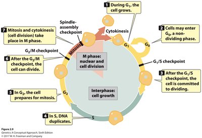
The cell cycle is the series of events that a cell undergoes from one division to the next. It includes interphase (G1, S, G2) and the mitotic phase (M).
G1 phase: Cell growth and preparation for DNA synthesis.
S phase: DNA replication.
G2 phase: Preparation for mitosis.
M phase: Mitosis and cytokinesis.
G0 phase: Non-dividing state.
Regulation of the Cell Cycle
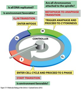
Cell cycle checkpoints ensure the fidelity of cell division by monitoring and regulating the progression through critical phases.
G1/S checkpoint: Checks for DNA damage before replication.
G2/M checkpoint: Ensures DNA replication is complete and undamaged before mitosis.
Spindle checkpoint (Metaphase-to-Anaphase): Ensures all chromosomes are properly attached to the spindle before chromatid separation.
Stages of Mitosis
Prophase: Chromosomes condense, spindle apparatus begins to form.
Prometaphase: Nuclear envelope breaks down, spindle formation completes, chromosomes attach to spindle via kinetochores.
Metaphase: Chromosomes align at the metaphase plate; spindle checkpoint ensures proper attachment.
Anaphase: Cohesins are degraded, sister chromatids separate and move to opposite poles.
Telophase: Chromatids reach poles, nuclear envelope reforms, spindle disassembles.
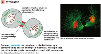
Cytokinesis: Division of cytoplasm, forming two independent daughter cells.
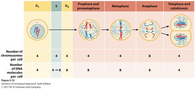
Summary Table: Chromosome and DNA Content During Mitosis
Stage | Number of Chromosomes per Cell | Number of DNA Molecules per Cell |
|---|---|---|
G1 | 4 | 4 |
S | 4 | 4 → 8 |
G2 | 4 | 8 |
Prophase/Prometaphase | 4 | 8 |
Metaphase | 4 | 8 |
Anaphase | 8 | 8 |
Telophase/Cytokinesis | 4 | 4 |
Meiosis
Purpose and Overview
Meiosis is a specialized form of cell division that reduces the chromosome number by half, producing four genetically unique haploid cells (gametes). It is essential for sexual reproduction and genetic diversity.
Consists of two sequential divisions: Meiosis I (reductional) and Meiosis II (equational).
Introduces genetic variation through recombination and independent assortment.
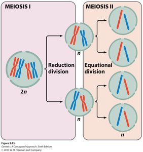
Stages of Meiosis I
Prophase I: Chromosomes condense, homologous chromosomes pair (synapsis), crossing over (recombination) occurs, nuclear envelope breaks down, spindle forms.
Metaphase I: Homologous pairs align at the metaphase plate, spindle fibers attach to kinetochores.
Anaphase I: Homologous chromosomes separate to opposite poles; sister chromatids remain attached.
Telophase I: Spindle disassembles, nuclear envelope reforms, cytokinesis may occur.
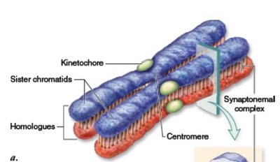
Prophase I Substages
Leptotene: Chromosomes begin to condense.
Zygotene: Homologous chromosomes pair via the synaptonemal complex (synapsis).
Pachytene: Synapsis complete, crossing over (recombination) occurs between non-sister chromatids.
Diplotene: Synaptonemal complex breaks down, homologs remain attached at chiasmata.
Diakinesis: Chromosomes further condense, nuclear envelope breaks down, spindle forms.
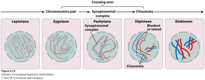
Homologous Chromosomes and Genetic Recombination
Homologous chromosomes: Chromosomes similar in shape and sequence, one from each parent.
Genetic recombination: Exchange of DNA between non-sister chromatids during crossing over, increasing genetic diversity.
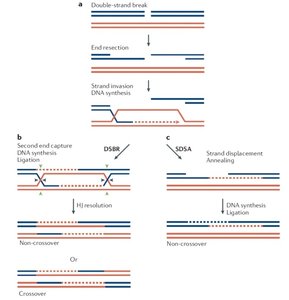
Mechanism of DNA Recombination: Holliday Model
Homologous chromosomes align, single-strand breaks occur, and strand invasion leads to the formation of Holliday junctions.
Branch migration and resolution of Holliday junctions (horizontal or vertical cleavage) result in crossover or non-crossover products.
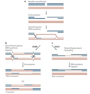
Stages of Meiosis II
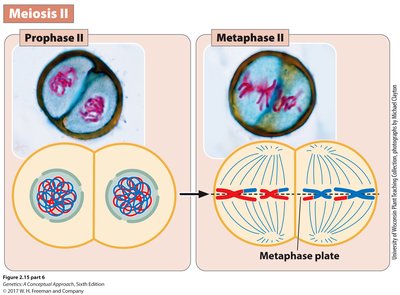
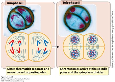
Meiosis II is similar to mitosis but occurs without prior DNA replication.
Phases: Prophase II, Metaphase II, Anaphase II, Telophase II, Cytokinesis.
Sister chromatids separate, resulting in four haploid cells.
Genetic Variation: Independent Assortment
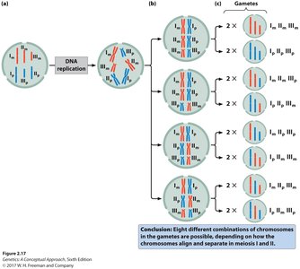
During metaphase I, the random alignment of homologous chromosome pairs leads to different combinations of chromosomes in gametes, further increasing genetic diversity.
Comparison of Mitosis, Meiosis I, and Meiosis II
Event | Mitosis | Meiosis I | Meiosis II |
|---|---|---|---|
Cell division | Yes | Yes | Yes |
Reduction in chromosome number | No | Yes | No |
Genetic variation produced | No | Yes | No |
Metaphase | Individual chromosomes line up | Homologous pairs line up | Individual chromosomes line up |
Anaphase | Chromatids separate | Homologous chromosomes separate | Chromatids separate |
Summary
Chromatin is less condensed DNA-protein complex; chromosomes are highly condensed forms visible during cell division.
Centromeres and telomeres are essential chromosome regions for segregation and stability.
DNA replication doubles the DNA content but not chromosome number until division occurs.
Binary fission, mitosis, and meiosis are distinct mechanisms of cell division with different outcomes.
Mitosis produces genetically identical cells; meiosis produces genetically diverse gametes.
Meiosis introduces genetic variation through recombination and independent assortment.
