 Indietro
DNA Replication: Mechanisms, Enzymes, and Structural Features
Guida di studio - Note intelligenti
 Appunti personalizzati basati sui tuoi materiali, ampliati con definizioni chiave, esempi e contesto.
DNA Replication
Main Features of the DNA Double Helix
The structure of DNA is fundamental to its function as the genetic material. The double helix model, proposed by Watson and Crick, describes two antiparallel strands held together by complementary base pairing.
Antiparallel Strands: The two DNA strands run in opposite directions (5' to 3' and 3' to 5').
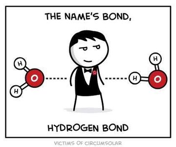
Base Pairing: Adenine (A) pairs with Thymine (T) via two hydrogen bonds, and Guanine (G) pairs with Cytosine (C) via three hydrogen bonds.
Hydrogen Bonds: These weak bonds allow the strands to separate during replication.
Helical Structure: The double helix is stabilized by base stacking interactions and the sugar-phosphate backbone.
Clues to the Replication of DNA
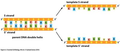
The specific pairing of bases in the DNA double helix immediately suggested a copying mechanism for genetic material. Each strand serves as a template for the synthesis of a new complementary strand.
Template Mechanism: Each parental DNA strand directs the synthesis of a new strand by complementary base pairing.
Semiconservative Replication: Each daughter DNA molecule consists of one parental and one newly synthesized strand.
Determining the Mechanism of DNA Replication
The Meselson-Stahl Experiment
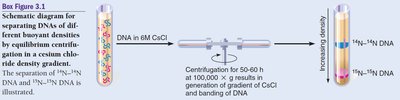
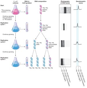
The Meselson-Stahl experiment (1958) provided definitive evidence for the semiconservative model of DNA replication using isotopic labeling and density gradient centrifugation.
Radioisotopes: DNA was labeled with heavy (15N) and light (14N) nitrogen isotopes to distinguish old and new DNA strands.
Centrifugation: High-speed centrifugation in a cesium chloride (CsCl) gradient separated DNA molecules by density.
Results: After one replication cycle, DNA had intermediate density, consistent with semiconservative replication. Further cycles produced both intermediate and light DNA bands.
Mechanism of DNA Replication
Directionality of DNA Synthesis
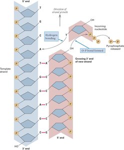
DNA polymerases can only add nucleotides to the 3' end of a growing DNA strand, so DNA synthesis always proceeds in the 5' to 3' direction.
5' to 3' Elongation: New nucleotides are added to the free 3'-OH group of the growing strand.
Complementary Base Pairing: Ensures accurate copying of genetic information (A pairs with T, G pairs with C).
Proteins Involved in DNA Replication
DNA replication is a highly coordinated process involving multiple enzymes and proteins, each with a specific role.
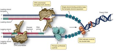
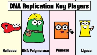
Helicase: Unwinds the DNA double helix at the replication fork.
Single-Strand Binding Proteins (SSBs): Stabilize unwound DNA strands.
Primase: Synthesizes short RNA primers to provide a starting point for DNA polymerase.
DNA Polymerase III: Main enzyme for synthesizing new DNA strands.
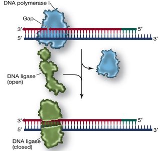
DNA Polymerase I: Removes RNA primers and fills in the gaps with DNA.
DNA Ligase: Joins Okazaki fragments on the lagging strand by forming phosphodiester bonds.
Leading vs. Lagging Strands
Because DNA polymerase can only synthesize DNA in the 5' to 3' direction, replication is continuous on one strand (leading) and discontinuous on the other (lagging).
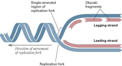
Leading Strand: Synthesized continuously in the direction of the replication fork.
Lagging Strand: Synthesized discontinuously as short Okazaki fragments, which are later joined by DNA ligase.
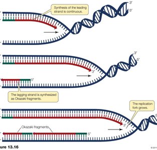
Okazaki Fragments and DNA Ligase
On the lagging strand, DNA is synthesized in short segments called Okazaki fragments. These fragments are joined together by DNA ligase after RNA primers are removed and replaced with DNA.
Okazaki Fragments: Short DNA segments synthesized on the lagging strand.
DNA Polymerase I: Removes RNA primers and fills in the gaps with DNA nucleotides.
DNA Ligase: Seals the nicks between Okazaki fragments, forming a continuous DNA strand.
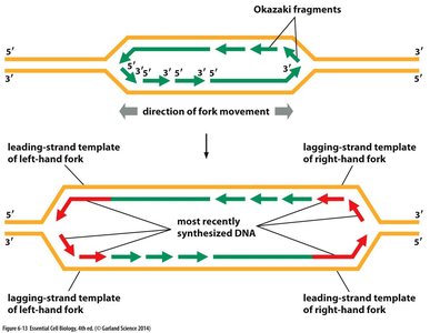
Replication Fork and Bidirectional Replication
Replication begins at origins of replication and proceeds bidirectionally, forming replication forks that move away from the origin.
Replication Fork: The Y-shaped region where DNA is unwound and new strands are synthesized.
Bidirectional Replication: Replication proceeds in both directions from each origin, increasing efficiency.
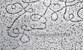
Supercoiling and DNA Structure
Supercoiling of DNA
Supercoiling refers to the overwinding or underwinding of DNA, which occurs in closed circular DNA molecules. Supercoiling compacts DNA and affects its accessibility for replication and transcription.
Definition: Supercoiling is the coiling of a closed duplex DNA in space so that it crosses over its own axis.
Function: Helps compact DNA and can regulate access to genetic information.
Summary Table: Key Enzymes in DNA Replication
Enzyme/Protein | Function |
|---|---|
Helicase | Unwinds the DNA double helix |
Single-Strand Binding Proteins | Stabilize unwound DNA |
Primase | Synthesizes RNA primers |
DNA Polymerase III | Main enzyme for DNA synthesis |
DNA Polymerase I | Removes RNA primers, fills gaps with DNA |
DNA Ligase | Joins Okazaki fragments |
Practice Question
If DNA ligase was missing from an in vitro DNA replication reaction, but all the other enzymes were available, you would expect to see:
A) Only the “old” DNA, no new DNA
B) Incorporation of incorrect nucleotides
C) Too many short fragments (Correct answer: Okazaki fragments would not be joined)
D) No Change
