 Indietro
Foundations of Molecular Genetics & the Central Dogma: Core Concepts and Clinical Applications
Guida di studio - Note intelligenti
 Appunti personalizzati basati sui tuoi materiali, ampliati con definizioni chiave, esempi e contesto.
Introduction to Molecular Genetics
Overview of Genetics and the Central Dogma
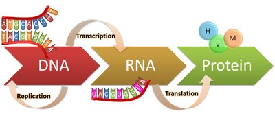
Genetics is the study of heredity and the variation of inherited characteristics. The central dogma of molecular biology describes the flow of genetic information within a biological system, outlining how DNA is transcribed into RNA and then translated into protein. This foundational concept is essential for understanding gene expression, mutation, and inheritance.
Genetic Terminology: Key terms include gene, genome, chromosome, allele, genotype, and phenotype.
Central Dogma: The process by which genetic information is transferred from DNA to RNA (transcription) and from RNA to protein (translation).
Clinical Relevance: Mutations in genetic material can lead to diseases by altering protein function.
DNA and Chromosome Structure
DNA Structure and Properties
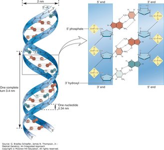
DNA (deoxyribonucleic acid) is a double-helical molecule composed of two antiparallel strands of nucleotides. Each nucleotide consists of a phosphate group, a deoxyribose sugar, and a nitrogenous base (adenine, thymine, guanine, or cytosine). The strands are held together by hydrogen bonds between complementary bases (A:T and G:C).
Antiparallel Strands: DNA strands run in opposite directions, with distinct 5’ and 3’ ends.
Phosphate-Sugar Backbone: Provides structural stability and a negative charge.
Base Pairing: Adenine pairs with thymine (A:T), and guanine pairs with cytosine (G:C).
Gene Structure
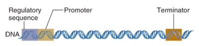
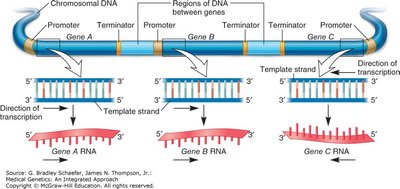
Genes are segments of DNA that encode functional products, typically proteins. Each gene contains regulatory regions, a promoter, coding regions (exons and introns), and a terminator sequence.
Promoter: Site where RNA polymerase binds to initiate transcription.
Regulatory Sequences: Control gene expression by interacting with transcription factors.
Coding Region: Contains exons (expressed sequences) and introns (intervening sequences).
Terminator: Signals the end of transcription.
Operons and Gene Regulation in Prokaryotes
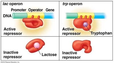
Operons are clusters of genes under the control of a single promoter, commonly found in prokaryotes. They allow coordinated regulation of genes with related functions.
Lac Operon: An inducible operon activated in the presence of lactose, allowing the expression of genes required for lactose metabolism.
Trp Operon: A repressible operon turned off in the presence of tryptophan, preventing unnecessary synthesis of tryptophan.
Genetic Information Flow: Replication, Transcription, and Translation
DNA Replication
DNA replication is a semi-conservative process in which each new DNA molecule consists of one original strand and one newly synthesized strand. Replication proceeds in the 5’ to 3’ direction, with new nucleotides added to the free 3’ hydroxyl group.
Semi-Conservative: Each daughter DNA contains one parental and one new strand.
Directionality: DNA polymerase synthesizes DNA in the 5’ to 3’ direction.
Transcription
Transcription is the process by which the genetic code in DNA is copied into messenger RNA (mRNA). RNA polymerase binds to the promoter region, and transcription factors regulate the initiation of transcription.
Initiation: RNA polymerase and transcription factors assemble at the promoter.
Elongation: RNA polymerase synthesizes the RNA strand complementary to the DNA template.
Termination: Transcription ends at the terminator sequence.
Translation and the Genetic Code
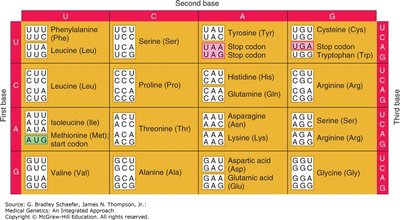
Translation is the process by which mRNA is decoded to synthesize proteins. Ribosomes read the mRNA sequence in codons (three-nucleotide sequences), each specifying an amino acid. The genetic code is universal and redundant, with multiple codons coding for the same amino acid.
Initiation: Ribosome assembles at the start codon (AUG).
Elongation: tRNAs bring amino acids to the ribosome, matching codons with anticodons.
Termination: Translation ends at a stop codon (UAA, UAG, UGA).
Chromosome Structure and Human Genome
Chromosome Organization
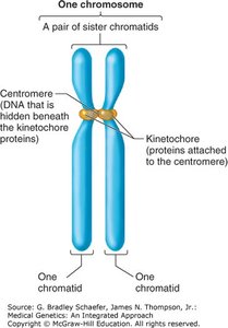
Chromosomes are structures composed of DNA and proteins (histones) that package and organize genetic material. Each chromosome contains a centromere, telomeres, and hundreds of genes.
Centromere: Central region important for chromosome segregation during cell division.
Telomeres: Repetitive sequences at chromosome ends, protecting against degradation.
Sister Chromatids: Identical copies formed after DNA replication, joined at the centromere.
Human Karyotype and Cytogenetic Nomenclature
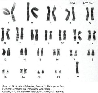
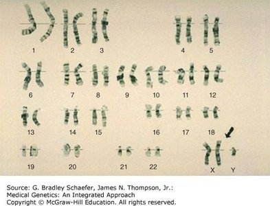
The normal human genome consists of 46 chromosomes: 22 pairs of autosomes and one pair of sex chromosomes. Karyotypes are annotated by total chromosome number, sex chromosomes, and any abnormalities (e.g., 46, XX; 47, XX, +21).
Autosomes: Non-sex chromosomes (pairs 1-22).
Sex Chromosomes: X and Y chromosomes determine biological sex.
Cytogenetic Notation: Used to describe chromosomal abnormalities (e.g., deletions, duplications).
Mutations and Chromosomal Aberrations
Types of Gene Mutations
Mutations are changes in the DNA sequence that can affect gene function. They are classified based on their nature and effect on the genetic code.
Frameshift Mutations: Insertions or deletions that alter the reading frame (except multiples of three nucleotides).
Substitution Mutations: Replacement of one nucleotide with another, which can be silent, missense, or nonsense (introducing a stop codon).
Chromosomal Aberrations
Chromosomal abnormalities involve changes in chromosome number or structure, often leading to clinical disorders.
Aneuploidy: Abnormal number of chromosomes (e.g., trisomy 21 in Down syndrome).
Polyploidy: More than two complete sets of chromosomes.
Structural Abnormalities: Deletions, duplications, inversions, and translocations.

Clinical Impact of Mutations
Mutations can disrupt metabolic pathways, leading to genetic disorders. For example, mutations in enzymes involved in phenylalanine metabolism can cause diseases such as phenylketonuria. Mutations in DNA repair genes (e.g., p53, BRCA1/2) increase cancer risk due to impaired genomic stability.
Tumor Suppressor Genes: Genes like p53 and Rb regulate cell cycle checkpoints and apoptosis.
DNA Repair Genes: Maintain genetic integrity; mutations increase mutation rates and cancer risk.
Cell Division: Mitosis and Meiosis
Cell Cycle and Checkpoints
The cell cycle consists of interphase (G1, S, G2) and mitotic phase (M). Checkpoints ensure proper cell division by monitoring DNA integrity, replication, and spindle attachment.
G1 Checkpoint: Checks for nutrients and DNA damage.
G2 Checkpoint: Ensures DNA replication is complete and undamaged.
M Checkpoint: Verifies spindle attachment before chromosome separation.
Mitosis
Mitosis is the process by which a cell divides to produce two genetically identical daughter cells. It consists of several phases: interphase, prophase, prometaphase, metaphase, anaphase, and telophase/cytokinesis.
Purpose: Growth, repair, and asexual reproduction.
Outcome: Two diploid cells identical to the parent cell.

Meiosis
Meiosis is a specialized form of cell division that produces four genetically unique haploid gametes. It involves two rounds of division (meiosis I and II) and introduces genetic variation through crossing over and independent assortment.
Purpose: Sexual reproduction and genetic diversity.
Outcome: Four haploid cells, each genetically distinct from the parent and each other.
Key Events: Homologous chromosomes separate in meiosis I; sister chromatids separate in meiosis II; crossing over occurs in prophase I.

Nondisjunction and Aneuploidy
Nondisjunction is the failure of chromosomes or chromatids to separate properly during meiosis, resulting in gametes with abnormal chromosome numbers. This can lead to aneuploid conditions such as Turner syndrome (45, XO) and Klinefelter syndrome (47, XXY).
Meiosis I Nondisjunction: Homologous chromosomes fail to separate; all resulting gametes are abnormal.
Meiosis II Nondisjunction: Sister chromatids fail to separate; half of the gametes are abnormal.
Sex Chromosome Aneuploidy: Conditions like Turner syndrome and Klinefelter syndrome are better tolerated than autosomal aneuploidies.
Summary of Key Concepts
Genetic information flows from DNA to RNA to protein via the central dogma.
Gene structure includes regulatory regions, promoters, coding sequences, and terminators.
Mutations and chromosomal aberrations can have significant clinical consequences.
Mitosis and meiosis are distinct processes with unique roles in growth, repair, and reproduction.
Understanding these molecular mechanisms is essential for diagnosing and treating genetic diseases.
