 Indietro
Gene Expression: Translation – Structure and Function of Proteins, Genetic Code, and Translation Mechanisms
Guida di studio - Note intelligenti
 Appunti personalizzati basati sui tuoi materiali, ampliati con definizioni chiave, esempi e contesto.
Gene Expression: Translation
Proteins and Amino Acids
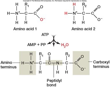
Proteins are essential macromolecules composed of amino acids, which are linked together by peptide bonds. Each amino acid contains an amino (NH2) group, a carboxyl (COOH) group, and a unique side chain (R group) that determines its properties. Amino acids are classified into four groups based on their side chains: acidic, basic, nonpolar, and polar (uncharged).
Amino acids: Building blocks of proteins, each with a unique R group.
Peptide bond: Covalent bond formed between the carboxyl group of one amino acid and the amino group of another, releasing water (H2O).
Protein structure: Determined by the sequence and chemical properties of amino acids.
Example: The formation of a peptide bond is a dehydration synthesis reaction.
Levels of Protein Structure
Proteins exhibit four levels of structural organization, each contributing to their function and stability.
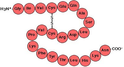
Primary structure: Linear sequence of amino acids in a polypeptide chain.
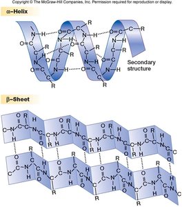
Secondary structure: Local folding patterns stabilized by hydrogen bonds, including alpha helices and beta sheets.
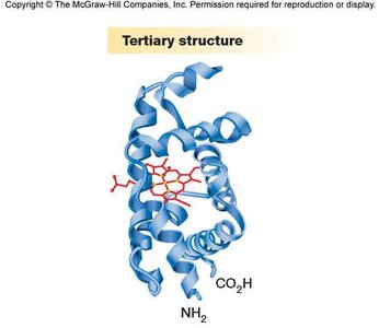
Tertiary structure: Overall three-dimensional shape of a single polypeptide, determined by interactions among side chains.
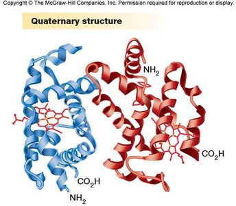
Quaternary structure: Association of multiple polypeptide subunits to form a functional protein.
Example: Hemoglobin consists of two alpha and two beta globin subunits.
The Genetic Code
Codons and Deciphering the Code
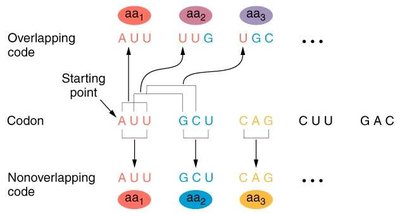
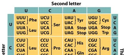
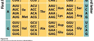
The genetic code is the set of rules by which information encoded in mRNA is translated into proteins. Each codon, a sequence of three nucleotides, specifies a particular amino acid. The code is non-overlapping and degenerate, meaning multiple codons can encode the same amino acid.
Codon: Three-nucleotide sequence in mRNA that specifies an amino acid.
Non-overlapping code: Each nucleotide is part of only one codon.
Degeneracy: Redundancy in the code; several codons may code for the same amino acid.
Example: UUU and UUC both code for phenylalanine.
Reading Frames and Mutations
The reading frame determines how the mRNA sequence is divided into codons. Insertions or deletions can shift the reading frame, resulting in frameshift mutations that alter the protein sequence.
Reading frame: The way nucleotides are grouped into codons for translation.
Frameshift mutation: Caused by insertion or deletion of nucleotides, changing the reading frame.
Example: Inserting or deleting a nucleotide in "PAT THE CAT" changes the grouping of letters.
tRNA Structure and Codon Recognition
tRNA Structure and Function
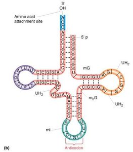
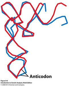
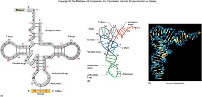
Transfer RNA (tRNA) molecules are responsible for bringing amino acids to the ribosome during translation. Each tRNA has a unique secondary structure, often described as a cloverleaf, and contains an anticodon that pairs with the codon in mRNA.
tRNA: Adaptor molecule that carries amino acids and recognizes codons via its anticodon.
Aminoacyl tRNA: tRNA charged with its corresponding amino acid.
Cloverleaf structure: Secondary structure of tRNA, positioning the anticodon for base pairing.
Example: tRNAPhe recognizes the UUU codon and carries phenylalanine.
Wobble Base Pairing and Modified Bases
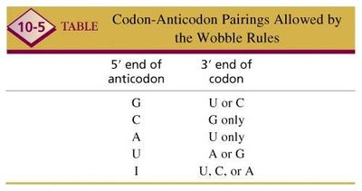
Wobble base pairing allows flexibility in the pairing between the third base of the codon and the first base of the anticodon, enabling fewer tRNAs to recognize multiple codons. Modified bases such as inosine contribute to this property.
Wobble: Non-standard base pairing at the third position of the codon, increasing efficiency.
Inosine (I): Modified base in tRNA that can pair with U, C, or A.
Example: In E. coli, 50 tRNAs can decode 61 sense codons due to wobble.
5' end of anticodon | 3' end of codon |
|---|---|
G | U or C |
C | G only |
A | U only |
U | A or G |
I | U, C, or A |
Start and Stop Codons, Universality, and Evolution of the Genetic Code
Start and Stop Codons
Translation begins at the start codon (AUG) and ends at one of the three stop codons (UAG, UAA, UGA), which do not code for amino acids.
Start codon: AUG (methionine)
Stop codons: UAG (amber), UAA (ochre), UGA (opal)
Nonsense codons: Another term for stop codons
Universality and Evolution of the Genetic Code
The genetic code is nearly universal among living organisms, with some exceptions in mitochondria and certain species. The code has evolved to minimize the impact of mutations, especially at the third position of codons.
Universality: Most organisms use the same genetic code.
Mutation minimization: Mutations in the third position often do not change the encoded amino acid.
Example: All codons with U as the middle base encode hydrophobic amino acids.
Translation Machinery and Process
Ribosomes and Translation Components
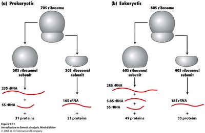
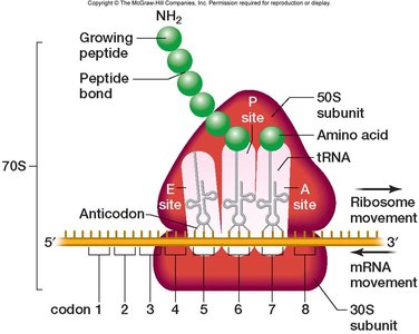
Translation occurs on ribosomes, which are composed of two subunits and contain binding sites for mRNA, tRNA, and accessory proteins. Ribosomes differ between prokaryotes and eukaryotes in size and composition.
Ribosome: Molecular machine for protein synthesis, composed of rRNA and proteins.
Subunits: Prokaryotic (30S + 50S = 70S), Eukaryotic (40S + 60S = 80S)
Binding sites: A (aminoacyl), P (peptidyl), and E (exit) sites for tRNA.
Charging tRNA: Aminoacyl-tRNA Synthetases
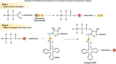
Aminoacyl-tRNA synthetases are enzymes that attach the correct amino acid to its corresponding tRNA. This charging process is essential for accurate translation.
Charging: Covalent attachment of amino acid to tRNA's 3' end.
Two-stage reaction: Amino acid activation (binds ATP), then transfer to tRNA.
Example: tRNA synthetase ensures fidelity in translation.
Translation: Steps and Mechanisms
Initiation (Prokaryotes)
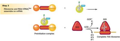
Translation initiation involves assembly of the ribosome on the mRNA, recognition of the start codon, and binding of the initiator tRNA. In prokaryotes, the Shine-Dalgarno sequence helps position the ribosome.
Initiator tRNA: Carries N-formyl methionine (fMet-tRNAMet).
Initiation factors: IF1, IF2, IF3 facilitate ribosome assembly.
Shine-Dalgarno sequence: Sequence in 5' UTR of mRNA that pairs with 16S rRNA.

Initiation (Eukaryotes)
In eukaryotes, translation initiation involves recognition of the 5' cap, scanning for the start codon, and assembly of the ribosome. The Kozak sequence surrounds the start codon to ensure correct initiation.
5' cap: Modified guanine nucleotide at the 5' end of mRNA.
Kozak sequence: Consensus sequence around AUG start codon.
Initiation factors: eIF2, eIF3, eIF4F, eIF5 facilitate assembly and scanning.
Elongation
Elongation is the process by which amino acids are added to the growing polypeptide chain. It involves the binding of charged tRNAs, peptide bond formation, and translocation of the ribosome.
Elongation factors: EF-Tu, EF-Ts, EF-G (prokaryotes); eEF1α, eEF1βγ, eEF2 (eukaryotes).
Peptidyl transferase: Ribosomal RNA enzyme that catalyzes peptide bond formation.
Translocation: Movement of the ribosome along mRNA by one codon.
Example: EF-Tu assists in tRNA binding; EF-G mediates translocation.
Termination
Translation terminates when a stop codon enters the A site. Release factors bind, the polypeptide is released, and the ribosome dissociates.
Release factors: Proteins that recognize stop codons and trigger release of the polypeptide.
Ribosomal recycling factor: Facilitates dissociation of ribosomal subunits.
Translation Efficiency, Energy Cost, and mRNA Types
Rate and Cost of Translation
Translation is a highly energy-intensive process, requiring multiple high-energy phosphate bonds for each amino acid added. Rates differ between prokaryotes and eukaryotes.
Prokaryotes: ~15 amino acids/second
Eukaryotes: ~5 amino acids/second
Energy cost: Four high-energy phosphate bonds per amino acid
Differences Between Prokaryotic and Eukaryotic mRNAs
Prokaryotic mRNAs are often polycistronic, encoding multiple proteins per transcript, while eukaryotic mRNAs are monocistronic, encoding a single protein.
Polycistronic: Multiple genes per mRNA (prokaryotes)
Monocistronic: One gene per mRNA (eukaryotes)
Specializations and Posttranslational Modifications
Targeting mRNA to the Endoplasmic Reticulum
Some eukaryotic proteins are synthesized on ribosomes bound to the endoplasmic reticulum (ER). Signal peptides direct the ribosome to the ER, where translation resumes and the protein is translocated into the ER lumen.
Signal peptide: Sequence of positively charged and hydrophobic amino acids
Signal Recognition Particle (SRP): Binds signal peptide and halts translation
Translocon: Channel in ER membrane for protein translocation
Signal peptidase: Removes signal peptide in ER
Posttranslational Changes to Proteins
After translation, proteins may undergo modifications such as folding, cleavage, phosphorylation, glycosylation, or lipid addition. Chaperones assist in proper folding.
Chaperones: Proteins that facilitate correct folding
Phosphorylation: Addition of phosphate groups
Glycosylation: Addition of sugars
Lipid addition: Attachment of lipid groups
Coupling of Transcription and Translation, Polysomes
Transcription and Translation Are Coupled in Prokaryotes
In prokaryotes, transcription and translation occur simultaneously, allowing rapid protein synthesis.
Polysomes
Multiple ribosomes can translate a single mRNA simultaneously, forming a polysome and increasing efficiency.
