 Indietro
Genetics Exam Study Guide: DNA Replication, Gene Regulation, and Protein Synthesis
Guida di studio - Note intelligenti
 Appunti personalizzati basati sui tuoi materiali, ampliati con definizioni chiave, esempi e contesto.
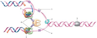
Q1. In the figure showing DNA replication in E. coli, what is the name of the protein labeled number 6?
Background
Topic: DNA Replication in Prokaryotes
This question tests your understanding of the proteins involved in the replication fork during DNA synthesis in E. coli. Knowing the roles of each protein is essential for understanding how DNA is copied accurately.
Key Terms and Concepts:
Replication fork: The area where the DNA is actively being unwound and replicated.
DNA polymerase III: The main enzyme responsible for synthesizing new DNA strands.
Sliding clamp (beta clamp): Helps DNA polymerase stay attached to the DNA.
SSB (Single-Stranded Binding protein): Stabilizes single-stranded DNA.
Step-by-Step Guidance
Examine the diagram and locate the protein labeled as number 6.
Recall the main enzymes and accessory proteins at the replication fork: DNA polymerase III, sliding clamp, SSB, primase, helicase, etc.
Consider the function of each protein and match it to the location and shape in the diagram.
Think about which protein is directly involved in synthesizing the new DNA strand on both the leading and lagging strands.
Try solving on your own before revealing the answer!
Final Answer: Pol III core enzyme
The protein labeled number 6 is the Pol III core enzyme, which is the main DNA polymerase responsible for synthesizing new DNA during replication in E. coli.
Q2. In the figure showing DNA replication in E. coli, what is the name of the protein labeled number 5?
Background
Topic: DNA Replication Fork Components
This question focuses on identifying the accessory protein that helps DNA polymerase maintain its association with the DNA template during replication.
Key Terms and Concepts:
Sliding clamp (beta clamp): A ring-shaped protein that encircles DNA and anchors DNA polymerase III to the template, increasing processivity.
Processivity: The ability of an enzyme to catalyze consecutive reactions without releasing its substrate.
Step-by-Step Guidance
Locate the protein labeled number 5 in the diagram.
Recall which protein forms a ring around DNA and is essential for keeping DNA polymerase attached during replication.
Compare the shape and position of number 5 to the known structure of the sliding clamp.
Think about the role of this protein in the context of the replication fork.
Try solving on your own before revealing the answer!
Final Answer: Sliding clamp (beta clamp)
The protein labeled number 5 is the sliding clamp (also known as the beta clamp), which helps DNA polymerase III stay attached to the DNA template during replication.
Q3. In the figure showing DNA replication in E. coli, what is the name of the protein labeled number 1?
Background
Topic: DNA Replication Fork Components
This question tests your knowledge of proteins that stabilize single-stranded DNA during replication, preventing it from re-annealing or being degraded.
Key Terms and Concepts:
SSB (Single-Stranded Binding protein): Binds to single-stranded DNA to protect it and keep it stable during replication.
DNA stability: Essential for accurate replication and preventing errors.
Step-by-Step Guidance
Find the protein labeled number 1 in the diagram.
Recall which protein binds to single-stranded DNA during replication.
Consider the function and location of SSB in the replication fork.
Match the label to the protein that stabilizes the unwound DNA.
Try solving on your own before revealing the answer!
Final Answer: SSB (Single-Stranded Binding protein)
The protein labeled number 1 is SSB, which binds to and stabilizes single-stranded DNA during replication.
Q4. DNA ligase is required to form phosphodiester linkages in nicks that are generated when DNA polymerase I removes an RNA primer and meets newly synthesized DNA ahead of it. This action seals the two strands together to form a continuous single strand.
Background
Topic: DNA Replication and Repair
This question tests your understanding of how DNA fragments are joined together during replication, especially on the lagging strand.
Key Terms and Concepts:
DNA ligase: An enzyme that catalyzes the formation of phosphodiester bonds between DNA fragments.
Phosphodiester linkage: The bond that connects the sugar-phosphate backbone of DNA.
Okazaki fragments: Short DNA fragments synthesized on the lagging strand.
Step-by-Step Guidance
Understand the process of lagging strand synthesis, which produces Okazaki fragments.
Recall that DNA polymerase I removes RNA primers and fills in the gaps with DNA.
Recognize that nicks remain between fragments after primer removal.
Identify the enzyme responsible for sealing these nicks to create a continuous DNA strand.
Try solving on your own before revealing the answer!
Final Answer: DNA ligase
DNA ligase is the enzyme that forms phosphodiester bonds between DNA fragments, sealing nicks and creating a continuous strand.
Q5. After initiation at a given point, unidirectional synthesis replicates strands in one direction only, whereas bidirectional synthesis replicates strands in both directions. Synthesis using the leading strand template proceeds in the direction of the replication fork, producing a long, unbroken complementary strand. It is said to be continuous. Synthesis using the lagging strand template proceeds in the direction opposite that of the replication fork and produces multiple, short fragments (Okazaki fragments) that must be ligated together to form a long complementary strand. This type of synthesis is said to be discontinuous.
Background
Topic: DNA Replication Mechanisms
This question tests your understanding of the directionality and continuity of DNA synthesis on the leading and lagging strands.
Key Terms and Concepts:
Bidirectional replication: DNA synthesis occurs in both directions from the origin.
Continuous synthesis: Occurs on the leading strand.
Discontinuous synthesis: Occurs on the lagging strand, producing Okazaki fragments.
Okazaki fragments: Short DNA segments synthesized on the lagging strand.
Step-by-Step Guidance
Recall the structure of the replication fork and the directionality of DNA synthesis.
Identify which strand is synthesized continuously and which is synthesized discontinuously.
Understand why Okazaki fragments are produced on the lagging strand.
Think about the enzymes involved in joining these fragments together.
Try solving on your own before revealing the answer!
Final Answer: Leading strand synthesis is continuous; lagging strand synthesis is discontinuous.
DNA replication is bidirectional, with the leading strand synthesized continuously and the lagging strand synthesized discontinuously as Okazaki fragments.
Q6. Alternative splicing occurs when copies of a pre-mRNA are spliced in more than one way, yielding various combinations of exons in the final mRNA products. Upon translation of a group of alternatively spliced mRNAs, a series of different, but related, proteins can be produced. What is the evolutionary advantage provided by alternative splicing?
Background
Topic: Post-transcriptional Regulation
This question tests your understanding of how alternative splicing increases protein diversity without increasing gene number.
Key Terms and Concepts:
Alternative splicing: The process by which different combinations of exons are joined to produce multiple mRNA variants from a single gene.
Exons: Coding regions of a gene.
Protein diversity: The variety of proteins produced by an organism.
Step-by-Step Guidance
Recall the process of splicing and how exons can be combined in different ways.
Think about how this affects the number of proteins produced from a single gene.
Consider the evolutionary implications of producing more proteins without increasing the genome size.
Try solving on your own before revealing the answer!
Final Answer: Alternative splicing allows organisms to encode more proteins without increasing gene number.
This increases protein diversity and functional complexity, providing an evolutionary advantage.
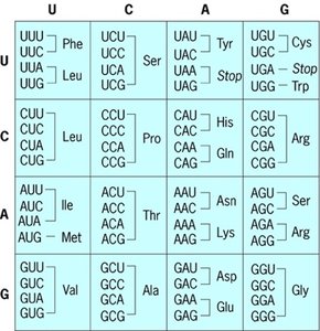
Q7. Predict the amino acid sequence (in one letter code) produced during translation of the short theoretical mRNA sequence: 5’-AUGCCGGAUUAUAGUUGA-3’
Background
Topic: Genetic Code and Translation
This question tests your ability to use the genetic code to translate an mRNA sequence into an amino acid sequence.
Key Terms and Concepts:
Codon: A sequence of three nucleotides in mRNA that specifies an amino acid.
Start codon (AUG): Codes for methionine and initiates translation.
Stop codon: Signals the end of translation.
One-letter amino acid code: Standard abbreviation for amino acids.
Step-by-Step Guidance
Divide the mRNA sequence into codons (groups of three nucleotides).
Use the genetic code table to match each codon to its corresponding amino acid.
Write out the amino acid sequence using the one-letter code.
Identify the stop codon and end the sequence there.
Try solving on your own before revealing the answer!
Final Answer: MPDYSG
The amino acid sequence is M (Methionine), P (Proline), D (Aspartate), Y (Tyrosine), S (Serine), and the sequence ends at the stop codon.
Q8. What would be the DNA sequence of the template strand that corresponds to the mRNA sequence (5’-AUGCCGGAUUAUAGUUGA-3’)?
Background
Topic: Transcription and DNA-mRNA Relationship
This question tests your understanding of how the template DNA strand is used to generate the mRNA sequence during transcription.
Key Terms and Concepts:
Template strand: The DNA strand used by RNA polymerase to synthesize mRNA.
Base pairing: A-U, G-C in RNA; A-T, G-C in DNA.
Directionality: mRNA is synthesized 5' to 3', so the template strand is read 3' to 5'.
Step-by-Step Guidance
Write the mRNA sequence and determine the complementary DNA sequence using base pairing rules.
Remember to replace uracil (U) in RNA with thymine (T) in DNA.
Write the DNA sequence in the 5' to 3' direction, matching the template strand.
Try solving on your own before revealing the answer!
Final Answer: 5’ TCAACTATAATCCGGCAT -3’
This is the DNA template strand that would produce the given mRNA sequence during transcription.
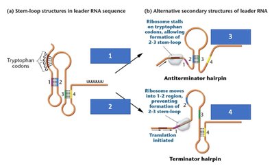
Q9. In the attenuation model for regulating the tryptophan operon, fill in the word or phrases that best describes what is in the 4 blue boxes in the diagram.
Background
Topic: Gene Regulation in Prokaryotes (Attenuation)
This question tests your understanding of how the trp operon is regulated by attenuation, a mechanism that uses leader RNA secondary structures to control transcription.
Key Terms and Concepts:
Attenuation: Regulation of gene expression by formation of alternative RNA secondary structures.
Leader sequence: Region of mRNA that can form stem-loop structures.
tRNATrp: tRNA charged with tryptophan.
Antiterminator and terminator hairpins: RNA structures that allow or prevent transcription.
Step-by-Step Guidance
Examine the diagram and identify the conditions for each box (charged tRNATrp available or not).
Recall how ribosome stalling or movement affects stem-loop formation.
Determine which hairpin forms and whether transcription proceeds or terminates.
Match the descriptions to the correct boxes in the diagram.
Try solving on your own before revealing the answer!
Final Answer:
Box 1: tRNATrp is NOT available
Box 2: tRNATrp is available
Box 3: Transcription proceeds
Box 4: Transcription terminates
These conditions determine whether the trp operon is transcribed or not, based on tryptophan availability.
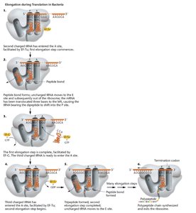
Q10. On a separate paper, write and draw out the process of elongation only in the translation process of proteins in prokaryotes. Do not write out the steps to initiation and termination!
Background
Topic: Translation Elongation in Prokaryotes
This question tests your ability to describe and illustrate the steps of elongation during protein synthesis, including the roles of mRNA, ribosome, tRNA, and elongation factors.
Key Terms and Concepts:
Elongation: The stage of translation where amino acids are added to the growing polypeptide chain.
Ribosome: The molecular machine that synthesizes proteins.
tRNA: Transfers amino acids to the ribosome.
Elongation factors: Proteins that facilitate the elongation process.
Step-by-Step Guidance
Describe the entry of charged tRNA into the A site of the ribosome.
Explain how peptide bond formation occurs between the amino acid in the A site and the growing chain in the P site.
Discuss the translocation of the ribosome, moving the tRNA from the A site to the P site.
Repeat the process for each new amino acid added to the chain.
Try solving on your own before revealing the answer!
Final Answer:
Elongation involves repeated cycles of tRNA entry, peptide bond formation, and ribosome translocation, resulting in the synthesis of a polypeptide chain. The diagram shows each step in detail.
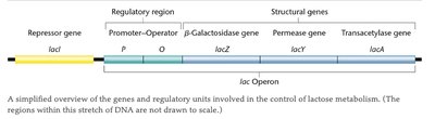
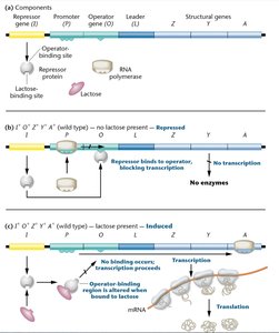
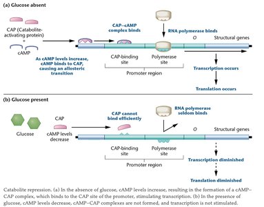
Q11. Describe how lactose metabolism in E. coli is regulated by an inducible system. Draw and label the lac operon, describe the function of the first two structural gene products, explain negative control, and explain positive control by CAP.
Background
Topic: Gene Regulation (lac Operon)
This question tests your understanding of the lac operon, including its structure, gene products, and regulatory mechanisms.
Key Terms and Concepts:
lac operon: A set of genes involved in lactose metabolism.
Inducible system: Gene expression is turned on in response to a substrate (lactose).
Negative control: Repressor protein blocks transcription in the absence of lactose.
Positive control: CAP-cAMP complex enhances transcription when glucose is absent.
β-galactosidase (lacZ): Converts lactose to glucose and galactose.
Permease (lacY): Facilitates lactose entry into the cell.
Step-by-Step Guidance
Draw and label the lac operon, including the regulatory region (lacI, promoter, operator) and structural genes (lacZ, lacY, lacA).
Describe the function of β-galactosidase (lacZ) and permease (lacY).
Explain how the repressor protein binds to the operator in the absence of lactose, preventing transcription (negative control).
Describe how the CAP-cAMP complex binds to the promoter in the absence of glucose, enhancing transcription (positive control).
Try solving on your own before revealing the answer!
Final Answer:
The lac operon is regulated by both negative and positive control mechanisms. In the absence of lactose, the repressor binds to the operator, blocking transcription. In the presence of lactose, the repressor is inactivated, allowing transcription. When glucose is absent, the CAP-cAMP complex binds to the promoter, enhancing transcription.
β-galactosidase (lacZ) converts lactose to glucose and galactose; permease (lacY) facilitates lactose entry into the cell.
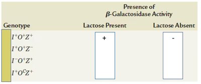
Q12. Examine the genotype table for the lac operon and provide the gene activity (+ or –) in the presence or absence of lactose for various E. coli genotypes.
Background
Topic: Gene Regulation (lac Operon Mutants)
This question tests your ability to predict gene expression based on different lac operon genotypes and environmental conditions.
Key Terms and Concepts:
lac operon genotypes: Variations in the regulatory and structural genes.
Gene activity: Expression of β-galactosidase (+ or –) depending on lactose presence.
Step-by-Step Guidance
Review the genotypes listed in the table.
Recall how the presence or absence of lactose affects gene expression for each genotype.
Fill in the table with + or – based on your understanding of the regulatory mechanisms.
Try solving on your own before revealing the answer!
Final Answer:
The gene activity is + in the presence of lactose and – in the absence of lactose for the wild-type genotype. For other genotypes, fill in the table based on the regulatory mutations and their effects.
