 Indietro
Genetics Study Guide: Introduction, Chromosomes, Mitosis, and Meiosis
Guida di studio - Note intelligenti
 Appunti personalizzati basati sui tuoi materiali, ampliati con definizioni chiave, esempi e contesto.
Introduction to Genetics
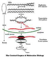
Central Dogma of Molecular Biology
The central dogma describes the flow of genetic information within a biological system. It explains how DNA is transcribed into RNA and then translated into protein, which determines cellular function and phenotype.
Replication: DNA duplicates itself to ensure genetic continuity.
Transcription: DNA is used as a template to synthesize RNA.
Translation: RNA is decoded to produce proteins, the functional molecules in cells.
Proteins: Carry out most cellular functions and determine traits.
Example: The process of gene expression, where a gene coding for an enzyme is transcribed and translated, resulting in the enzyme's production.
Key Terms in Genetics
Understanding genetics requires familiarity with foundational terminology.
Gene: Unit of heredity, usually codes for a polypeptide or RNA molecule.
Gene Expression: The process by which genetic information is accessed and used to synthesize proteins.
Trait: Observable characteristic resulting from gene expression.
Genotype: The genetic makeup of an individual.
Allele: Different forms of a gene.
Phenotype: The physical manifestation of the genotype.
Monohybrid Cross: Genetic cross involving one trait.
Dihybrid Cross: Genetic cross involving two traits.
True Breeding: Organisms that consistently produce the same phenotype over generations.
DNA, Chromosomes, Genes, and Proteins
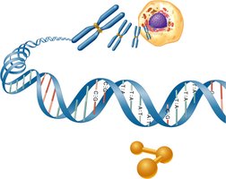
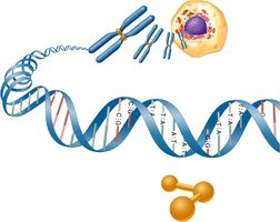
Genetic information is organized hierarchically from DNA to chromosomes, genes, and proteins. Each cell contains chromosomes made of DNA, which house genes that code for proteins.
DNA: The molecule of life, composed of nucleotide bases (A, T, C, G).
Chromosomes: Structures containing DNA and associated proteins.
Genes: Segments of DNA coding for proteins.
Proteins: Molecules that perform most cellular functions.
Example: Human cells contain 46 chromosomes, approximately 2 meters of DNA, and 20-25,000 genes.
Gene Expression
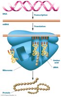
Gene expression involves transcription and translation, resulting in the synthesis of proteins from genetic information.
Transcription: DNA is converted to mRNA in the nucleus.
Translation: mRNA is decoded by ribosomes to synthesize proteins.
Example: The production of a pigment enzyme from a pigmentation gene.
Genotype Dictates Phenotype
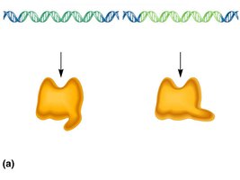
The genotype determines the phenotype through the process of gene expression. Different alleles can result in proteins with varying functionality, affecting observable traits.
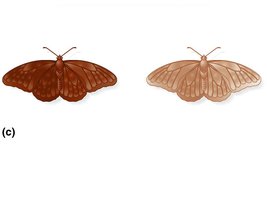
Highly functional enzyme: Results in a dark phenotype (e.g., dark butterfly).
Poorly functional enzyme: Results in a light phenotype (e.g., light butterfly).
Example: Variation in pigmentation due to different alleles of the pigmentation gene.
Chromosomes and Cell Division
Chromosome Structure and Function
Chromosomes are the carriers of genetic material in cells. Genes are located along chromosomes, which are composed of DNA, protein, and RNA. Chromatin is the form of DNA during interphase.
Chromosomes: Long molecules of DNA with associated proteins.
Chromatin: The less condensed form of DNA during interphase.
Gene Locus: The physical location of a gene on a chromosome.
Human Karyotype
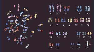
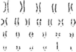
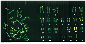
A karyotype is an organized profile of a person's chromosomes. Human somatic cells are diploid, containing two sets of chromosomes (one from each parent).
Diploid: Two sets of each chromosome.
Homologous Chromosomes: Chromosomes with the same genes but possibly different alleles.
Example: Human karyotype showing 23 pairs of chromosomes.
Chromosome Size and Shape
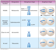
Chromosomes are classified based on the position of the centromere. The short arm is designated 'p' and the long arm 'q'.
Metacentric: Centromere in the middle.
Submetacentric: Centromere between middle and end.
Acrocentric: Centromere close to end.
Telocentric: Centromere at the end.
Centromere location | Designation | Metaphase shape | Anaphase shape |
|---|---|---|---|
Middle | Metacentric | Sister chromatids, centromere in center | Migration to poles |
Between middle and end | Submetacentric | p arm, q arm | Migration to poles |
Close to end | Acrocentric | Centromere near end | Migration to poles |
At end | Telocentric | Centromere at end | Migration to poles |
Homologous Chromosomes and Gene Loci
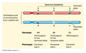
Homologous chromosomes have the same gene loci but may carry different alleles. The genotype is determined by the combination of alleles present.
Homozygous: Same allele at a locus.
Heterozygous: Different alleles at a locus.
Example: AA (homozygous dominant), Bb (heterozygous), cc (homozygous recessive).

Sex Determining Chromosomes
Sex chromosomes (X and Y in humans) determine biological sex and are not homologous but behave as homologs during meiosis.
X Chromosome: Contains many genes.
Y Chromosome: Contains fewer genes, determines male sex.
Example: Human XY karyotype.
Cell Division: Mitosis and Meiosis
Binary Fission in Prokaryotes
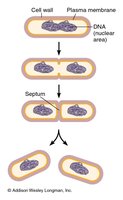
Prokaryotes divide by binary fission, a simple process involving the replication of a single circular chromosome followed by cell division.
Replication: Chromosome is duplicated.
Division: Each daughter cell receives one chromosome.
Eukaryotic Chromosomes and Cell Cycle
Eukaryotic cells have multiple linear chromosomes and divide by mitosis or meiosis. The cell cycle consists of interphase (G1, S, G2) and mitosis.
Interphase: Cell growth and DNA replication.
Mitosis: Division of the nucleus.
Cytokinesis: Division of the cytoplasm.
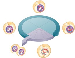
Stages of Mitosis
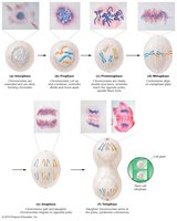
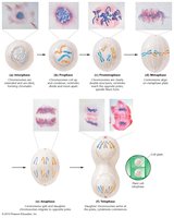
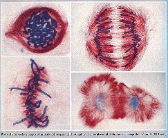
Mitosis is the process by which somatic cells divide, resulting in two genetically identical daughter cells. The stages include prophase, prometaphase, metaphase, anaphase, and telophase.
Prophase: Chromatin condenses into chromosomes.
Prometaphase: Nuclear envelope breaks down, spindle forms.
Metaphase: Chromosomes align at the metaphase plate.
Anaphase: Sister chromatids are pulled apart.
Telophase: Nuclear envelope reforms, cytokinesis occurs.
Chromosome Movement in Mitosis
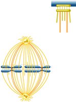
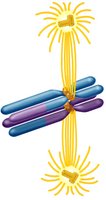
Microtubules attach to chromosomes at the kinetochore and pull them apart during mitosis.
Kinetochore: Protein structure on the centromere where microtubules attach.
Metaphase Plate: Area where chromosomes align before separation.
Mitosis in Plants
Plant cells undergo mitosis similar to animal cells, with the formation of a cell plate during cytokinesis.
Meiosis: Formation of Haploid Gametes
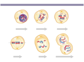
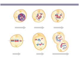
Meiosis is a specialized cell division process that produces haploid gametes for sexual reproduction. It involves one round of DNA replication and two rounds of cell division (Meiosis I and II).
Meiosis I: Homologous chromosomes separate, cells become haploid.
Meiosis II: Sister chromatids separate, resulting in four haploid cells.
Genetic Variation: Increased by crossing over and independent assortment.
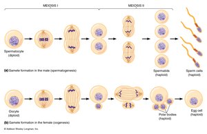
Products of Meiosis in Animals
Meiosis results in four haploid cells, which develop into gametes (sperm or egg cells).
Spermatogenesis: Formation of sperm cells.
Oogenesis: Formation of egg cells.
Comparison: Mitosis vs. Meiosis
Mitosis and meiosis are distinct processes with different outcomes and purposes.
Mitosis: Produces two genetically identical cells for growth and repair.
Meiosis: Produces four genetically diverse haploid cells for sexual reproduction.
Nondisjunction: Failure of chromosome separation, leading to mutations.
Summary Table: Mitosis vs. Meiosis
Process | Number of Divisions | Resulting Cells | Genetic Identity | Purpose |
|---|---|---|---|---|
Mitosis | 1 | 2 | Identical | Growth, repair |
Meiosis | 2 | 4 | Diverse | Sexual reproduction |
Additional info:
Genetic variation is crucial for evolution and adaptation in populations.
Chromosome abnormalities can lead to genetic disorders.
