 Indietro
Primary Mitochondrial Disorders: Genetic Basis, Clinical Features, and Diagnostic Strategies
Guida di studio - Note intelligenti
 Appunti personalizzati basati sui tuoi materiali, ampliati con definizioni chiave, esempi e contesto.
Primary Mitochondrial Disorders
Overview
Primary mitochondrial disorders are a diverse group of genetic diseases caused by dysfunction of the mitochondrial respiratory chain, the essential pathway for aerobic metabolism. These disorders can affect multiple organ systems, especially those with high energy demands, and are caused by pathogenic variants in either mitochondrial DNA (mtDNA) or nuclear DNA (nDNA) genes encoding mitochondrial proteins.
Genetic Basis of Mitochondrial Disorders
Mitochondrial DNA (mtDNA)
The human mitochondrial genome is a circular DNA molecule encoding essential components for oxidative phosphorylation. It contains 37 genes: 13 protein-coding genes, 22 tRNA genes, and 2 rRNA genes. The D-loop is a noncoding region involved in the regulation of transcription and replication.
Protein-coding genes: Encode subunits of respiratory chain complexes I, III, IV, and V.
tRNA and rRNA genes: Required for intra-mitochondrial protein synthesis.
Replication origins: OH (heavy strand) and OL (light strand).
Homoplasmy vs. Heteroplasmy: Homoplasmy refers to identical mtDNA copies in a cell; heteroplasmy is the coexistence of mutated and wild-type mtDNA, leading to variable clinical expression depending on the proportion of mutated mtDNA (threshold effect).
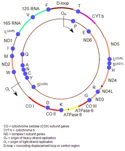
Figure: Structure of the human mitochondrial genome, showing gene locations and replication origins.
Nuclear DNA (nDNA)
Most mitochondrial proteins are encoded by nuclear genes. These genes are responsible for:
Maintenance and replication of mtDNA
Mitochondrial protein synthesis
Assembly and function of respiratory chain complexes
Coenzyme Q10 biosynthesis
Mitochondrial structure
Clinical Manifestations of Mitochondrial Disorders
General Features
Mitochondrial disorders can affect a single organ or multiple systems, often presenting with neurologic and muscular symptoms. Onset can occur at any age, and clinical features are highly variable due to heteroplasmy and tissue distribution of mutated mtDNA.
Common features: Ptosis, external ophthalmoplegia, myopathy, exercise intolerance, cardiomyopathy, sensorineural deafness, optic atrophy, pigmentary retinopathy, diabetes mellitus.
Central nervous system: Encephalopathy, seizures, dementia, migraine, stroke-like episodes, ataxia, spasticity.
Other: Increased risk of pregnancy loss.
Clinical Syndromes of mtDNA Disorders
Many mtDNA disorders present as distinct clinical syndromes. The following table summarizes key syndromes and their features:
Disorder | Primary Features | Additional Features |
|---|---|---|
CPEO | External ophthalmoplegia, bilateral ptosis, mild proximal myopathy | |
KSS | PEO onset <20 yrs, pigmentary retinopathy, CSF protein >1 g/L or cerebellar ataxia or heart block | Bilateral deafness, myopathy, dysphagia, diabetes mellitus, hypoparathyroidism, dementia |
Pearson syndrome | Sideroblastic anemia, pancytopenia, exocrine pancreatic failure | Renal tubular defects |
Leigh syndrome | Subacute relapsing encephalopathy, cerebellar/brain stem signs, infantile onset | Basal ganglia lucencies, maternal history of neurologic disease |
NARP | Peripheral neuropathy, ataxia, pigmentary retinopathy | Basal ganglia lucencies, abnormal electroretinogram, sensorimotor neuropathy |
MELAS | Stroke-like episodes <40 yrs, seizures/dementia, ragged-red fibers/lactic acidosis | Diabetes, cardiomyopathy, deafness, pigmentary retinopathy, cerebellar ataxia |
MERRF | Myoclonus, seizures, cerebellar ataxia, myopathy | Dementia, optic atrophy, deafness, neuropathy, spasticity, lipomata |
LHON | Bilateral visual failure, males:females ~4:1, median onset 24 yrs | Dystonia, cardiac pre-excitation syndromes |
Abbreviations: CPEO = chronic progressive external ophthalmoplegia; KSS = Kearns-Sayre syndrome; LHON = Leber hereditary optic neuropathy; MELAS = mitochondrial encephalomyopathy with lactic acidosis and stroke-like episodes; MERRF = myoclonic epilepsy with ragged-red fibers; NARP = neurogenic weakness with ataxia and retinitis pigmentosa.
Nuclear DNA Mitochondrial Disorders
nDNA mitochondrial disorders are clinically variable and may not fit classic syndromes. They can be classified by the affected mechanism, such as respiratory chain subunits, assembly factors, translation factors, or disorders associated with multiple mtDNA deletions or depletion.
Examples: Leigh syndrome (various complex deficiencies), hepatopathy, cardiomyopathy, ataxia neuropathy syndromes, coenzyme Q10 deficiency, Barth syndrome.
Diagnosis and Evaluation Strategies
Clinical Evaluation
Comprehensive physical and neurologic examination to identify syndromic features and organ involvement.
Family history (three generations) to assess inheritance patterns (maternal, autosomal dominant/recessive, X-linked).
Laboratory and Imaging Studies
Plasma/CSF lactate: Elevated levels support diagnosis but normal values do not exclude disease.
Neuroimaging: MRI/CT may show basal ganglia calcification, atrophy, or leukoencephalopathy.
Neurophysiology: EEG, EMG, nerve conduction studies for neurologic involvement.
Other: Cardiac evaluation, glucose testing for diabetes, muscle biopsy for respiratory chain function.
Molecular Genetic Testing
Targeted single-gene or multigene panel testing based on clinical suspicion.
Comprehensive genomic testing (exome/genome sequencing) for broad analysis.
Consider tissue selection for mtDNA analysis due to heteroplasmy (e.g., muscle or urinary epithelium).
Genetic Counseling and Inheritance Patterns
Modes of Inheritance
mtDNA mutations: Maternal inheritance; all children of affected mothers are at risk, but not those of affected fathers.
nDNA mutations: Autosomal recessive, autosomal dominant, or X-linked inheritance possible.
Risk to Family Members
mtDNA: Risk to siblings and offspring depends on maternal status and heteroplasmy level.
Autosomal recessive: 25% risk to siblings if both parents are carriers.
Autosomal dominant: 50% risk to offspring if a parent is affected.
X-linked: Affected males transmit to all daughters (carriers or affected), not sons.
Prenatal Testing
Prenatal diagnosis for mtDNA disorders is challenging due to heteroplasmy and tissue distribution.
nDNA mutations: Prenatal and preimplantation genetic testing possible once familial variants are identified.
Patient Care Guidelines
Regular surveillance for complications (cardiac, diabetes, neurologic).
Management is supportive; no curative treatments currently available.
Idebenone is approved for Leber hereditary optic neuropathy (LHON) in some countries.
Summary Table: Key Features of Primary Mitochondrial Disorders
Aspect | mtDNA Disorders | nDNA Disorders |
|---|---|---|
Inheritance | Maternal | Autosomal recessive, dominant, or X-linked |
Clinical Onset | Any age; often variable | Any age; often childhood |
Clinical Features | Often syndromic (e.g., MELAS, LHON) | Variable, may overlap syndromes |
Diagnosis | mtDNA analysis (muscle, urine, blood) | nDNA gene panels, exome/genome sequencing |
Additional info: Mitochondrial disorders are a major topic in medical genetics, intersecting with molecular genetics, inheritance patterns, and clinical diagnosis. Understanding the dual genetic origin (mtDNA and nDNA) and the concept of heteroplasmy is essential for interpreting clinical variability and genetic counseling.
