 Indietro
The Molecular Basis of Heredity: Discovery of DNA Structure
Guida di studio - Note intelligenti
 Appunti personalizzati basati sui tuoi materiali, ampliati con definizioni chiave, esempi e contesto.
The Molecular Basis of Heredity
Historical Foundations and Key Techniques
The discovery of DNA's structure was a pivotal moment in genetics, relying on advances in physics, chemistry, and biology. This section explores the historical context, experimental techniques, and key contributors to our understanding of the molecular basis of heredity.
Electromagnetic Spectrum and X-rays
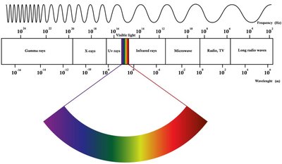
Electromagnetic Spectrum: The electromagnetic spectrum encompasses all types of electromagnetic radiation, including gamma rays, X-rays, ultraviolet (UV), visible light, infrared, microwaves, and radio waves.
X-rays: X-rays are a form of high-energy electromagnetic radiation, with wavelengths shorter than UV light but longer than gamma rays. They are particularly useful in probing the atomic structure of molecules.
Application in Genetics: X-rays are used in X-ray crystallography to determine the three-dimensional structure of biological macromolecules, such as DNA.
X-ray Diffraction and Crystallography
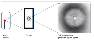
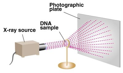
X-ray diffraction is a technique that allows scientists to infer the structure of crystalline substances by analyzing the pattern produced when X-rays are scattered by the atoms in a crystal.
Principle: When X-rays strike a crystal, they are diffracted in specific directions, creating a pattern that can be recorded and analyzed.
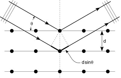
Bragg's Law: The relationship between the angle of incidence, the wavelength of X-rays, and the distance between crystal planes is given by Bragg's Law: where is an integer, is the wavelength, is the distance between planes, and is the angle of incidence.
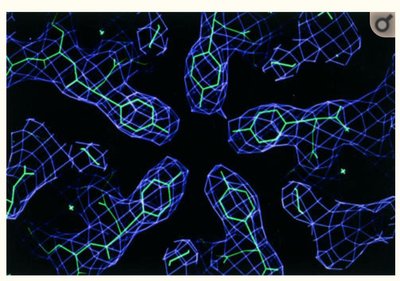
Electron Density Maps: The diffraction data are used to generate electron density maps, which reveal the positions of atoms within the molecule.
Discovery of the Double Helix Structure of DNA
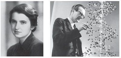
The elucidation of DNA's double helix structure was achieved through the integration of X-ray diffraction data and chemical knowledge. Key contributors included Rosalind Franklin, Maurice Wilkins, James Watson, and Francis Crick.
Key Experimental Evidence
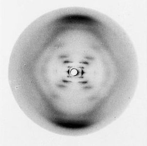
Rosalind Franklin & Maurice Wilkins: Their X-ray diffraction studies provided critical evidence that DNA is a helical molecule with regular structural features, including a 1 nm radius and a 3.4 nm helical pitch.
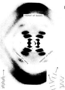
Photo 51: Franklin's famous X-ray diffraction image (Photo 51) revealed the helical nature and periodicity of DNA, which was instrumental in guiding Watson and Crick's model building.
Watson and Crick's Double Helix Model
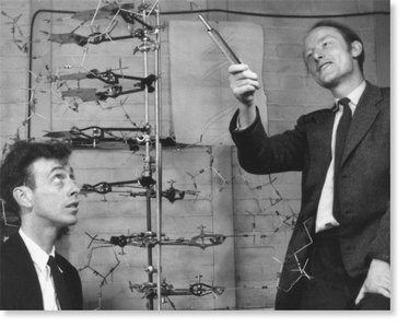
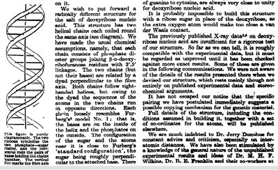
Model Proposal: In 1953, James Watson and Francis Crick proposed the double helix model of DNA, integrating data from X-ray diffraction and chemical analysis.
Key Features:
Two antiparallel polynucleotide chains coiled around a common axis.
Base pairing between adenine (A) and thymine (T), and between guanine (G) and cytosine (C), held together by hydrogen bonds.
Regular helical structure with a diameter of about 2 nm and a helical repeat every 3.4 nm (10 base pairs per turn).
Significance: The structure explained how genetic information could be stored, replicated, and transmitted.
Contributions of Rosalind Franklin
Biography: Rosalind Franklin (1920–1958) was a pioneering scientist whose expertise in X-ray crystallography was crucial to the discovery of DNA's structure.
Key Achievements:
Produced high-quality X-ray diffraction images of DNA, including Photo 51.
Her data provided essential measurements for the double helix model.
Continued research in structural biology until her untimely death at age 37.
Recognition: The Nobel Prize in Physiology or Medicine (1962) was awarded to Watson, Crick, and Wilkins for the discovery of the DNA structure. Franklin was not included, as the Nobel is not awarded posthumously.
Summary Table: Key Contributors and Techniques in DNA Structure Discovery
Scientist(s) | Contribution | Technique |
|---|---|---|
Rosalind Franklin | X-ray diffraction images (Photo 51), measurements of DNA dimensions | X-ray crystallography |
Maurice Wilkins | Collaboration on X-ray studies, provided data to Watson and Crick | X-ray crystallography |
James Watson & Francis Crick | Proposed the double helix model of DNA | Molecular modeling, integration of chemical and physical data |
Key Terms and Concepts
Double Helix: The three-dimensional structure of DNA, consisting of two intertwined strands forming a helical shape.
X-ray Crystallography: A technique for determining the atomic and molecular structure of a crystal by measuring the angles and intensities of X-rays diffracted by the crystal lattice.
Base Pairing: The specific hydrogen bonding between purines and pyrimidines (A-T and G-C) in DNA.
Electron Density Map: A three-dimensional representation of electron density within a molecule, derived from X-ray diffraction data.
Conclusion
The discovery of the DNA double helix was a landmark in genetics, made possible by the integration of X-ray crystallography, chemical analysis, and model building. The collaborative efforts of Franklin, Wilkins, Watson, and Crick laid the foundation for modern molecular genetics and our understanding of heredity.
