 Indietro
The Muscular System: Structure, Function, and Physiology
Guida di studio - Note intelligenti
 Appunti personalizzati basati sui tuoi materiali, ampliati con definizioni chiave, esempi e contesto.
The Muscular System
Introduction to Muscles
Muscle tissue is essential for movement and is present in every organ of the human body. Muscles account for a significant portion of body weight and are involved in both voluntary and involuntary activities.
Muscle tissue makes up about 40% of body weight in males and 32% in females.
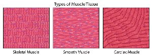
There are three main types of muscle tissue:
Skeletal muscle: Attaches to the skeleton, providing strength and mobility.
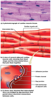
Cardiac muscle: Found exclusively in the heart.
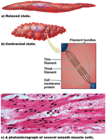
Smooth muscle: Located in the walls of the digestive tract, blood vessels, uterus, and ureters.
Muscle Attachment and Movement
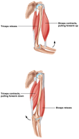
Muscles attach to bones via tendons, enabling movement of the skeleton. The coordinated contraction and relaxation of muscles allow for complex movements and maintenance of posture.
Tendons connect muscle to bone.
Muscles can be classified as:
Synergistic: Work together to produce the same movement.
Antagonistic: Oppose each other's actions.
Origin: The end of the muscle attached to a stationary bone.
Insertion: The end attached to a movable bone across a joint.
Muscle Structure: From Whole Muscle to Sarcomere
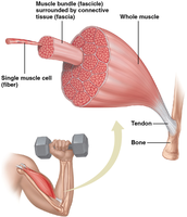
Muscles are organized in a hierarchical structure, from the whole muscle down to the contractile units called sarcomeres.
Muscle: Group of muscle cells with the same origin, insertion, and function.
Fascicles: Bundles of muscle fibers wrapped in connective tissue (fascia).
Muscle fibers (cells): Long, multinucleate cells packed with myofibrils.
Myofibrils: Cylindrical structures containing the proteins actin and myosin.
The Sarcomere: The Contractile Unit
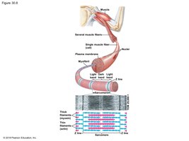
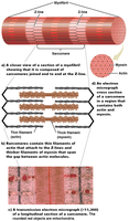
The sarcomere is the fundamental contractile unit of muscle, responsible for the striated appearance of skeletal muscle and the mechanism of contraction.
Sarcomere: Segment of a myofibril between two Z-lines.
Myosin: Forms thick filaments.
Actin: Forms thin filaments, attached to Z-lines.
During contraction, sarcomeres shorten as actin and myosin filaments slide past each other.
Muscle Contraction and Relaxation
Muscle contraction is initiated by nerve activation, which leads to a cascade of events resulting in the sliding of filaments within the sarcomere.
Muscle contraction: Each sarcomere shortens slightly.
Nerve activation increases calcium ion concentration near contractile proteins.
Calcium enables contraction; when stimulation stops, contraction ends.
Nerve Activation of Skeletal Muscle
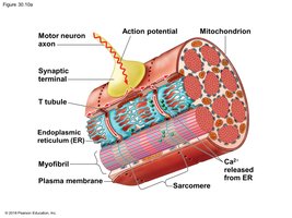
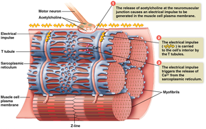
Motor neurons stimulate muscle contraction at the neuromuscular junction by releasing the neurotransmitter acetylcholine.
Acetylcholine binds to muscle cell receptors, generating an electrical impulse.
The impulse travels along T tubules, triggering calcium release from the sarcoplasmic reticulum.
Sliding Filament Mechanism
The sliding filament mechanism explains how muscles contract at the molecular level.
In resting muscle, myosin heads do not contact actin.
During contraction, myosin heads form cross-bridges with actin, bend, and pull actin filaments toward the center of the sarcomere.
This process repeats, shortening the sarcomere and the muscle.
Role of Calcium and Regulatory Proteins
Calcium ions play a critical role in initiating muscle contraction by interacting with regulatory proteins on actin filaments.
Troponin–tropomyosin complex blocks myosin binding sites on actin in the absence of calcium.
When calcium binds to troponin, the complex shifts, exposing binding sites and allowing cross-bridge formation.
Muscle contraction continues as long as calcium and ATP are present.
Muscle Relaxation
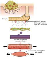
When nerve activation ends, calcium is pumped back into the sarcoplasmic reticulum, and the muscle relaxes.
ATP is required for both contraction and relaxation.
Without calcium, cross-bridges cannot form, and the muscle returns to its resting state.
Energy for Muscle Contraction
Muscle contraction requires significant energy, primarily in the form of ATP. Muscles have several mechanisms to produce and store ATP.
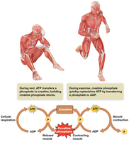
ATP is used to energize myosin heads and detach them from actin.
ATP is replenished by:
Creatine phosphate: Rapidly transfers a phosphate to ADP to form ATP.
Stored glycogen: Hydrolyzed to glucose for anaerobic metabolism.
Aerobic metabolism: Uses glucose, fatty acids, and other molecules.
Types of Muscle Contractions
Muscles can contract in different ways depending on the type of movement required.
Isotonic contractions: Muscle shortens while maintaining constant force; movement occurs.
Isometric contractions: Muscle tension increases, but the muscle does not shorten; no movement occurs.
Motor Units and Muscle Force
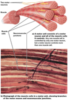
The force generated by a muscle depends on the number and size of motor units activated, as well as the frequency of stimulation.
Motor unit: A motor neuron and all the muscle cells it controls.
Larger motor units generate more force but less fine control (e.g., thigh muscles).
Smaller motor units allow for fine control (e.g., eye muscles).
Muscle tension is influenced by motor unit size, number of active units, and stimulation frequency.
Muscle Twitch, Summation, and Tetanus
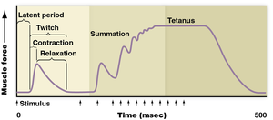
A muscle twitch is a single contraction-relaxation cycle in response to a stimulus. Increased frequency of stimulation leads to summation and, eventually, tetanus (sustained contraction).
Latent period: Delay between stimulus and contraction.
Summation: Increased force due to rapid, repeated stimulation.
Tetanus: Maximum, sustained contraction.
Slow-Twitch vs. Fast-Twitch Muscle Fibers
Muscle fibers are classified based on their contraction speed and metabolic pathways.
Slow-twitch fibers:
Contract slowly, use aerobic metabolism, many mitochondria, well-supplied with blood vessels, little glycogen, red color.
Used for endurance activities (e.g., jogging, swimming).
Fast-twitch fibers:
Contract quickly, use anaerobic metabolism, fewer mitochondria, less blood supply, store more glycogen, white color.
Used for brief, high-intensity activities (e.g., sprinting, lifting weights).
Exercise Training and Muscle Adaptation
Different types of exercise training lead to specific adaptations in muscle tissue.
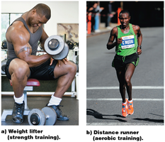
Strength training: Builds more myofibrils, increases muscle mass and strength (especially in fast-twitch fibers).
Aerobic training: Increases endurance, blood supply, mitochondria, and myoglobin in muscle cells.
Cardiac and Smooth Muscle
Cardiac and smooth muscles are involuntary and have unique structural and functional characteristics.
Cardiac muscle:
Found only in the heart, cells joined by intercalated discs with gap junctions for electrical connectivity.
Pacemaker cells set the rhythm of contraction.
Smooth muscle:
Found in walls of hollow organs, filaments arranged in criss-crossed bundles (no sarcomeres), no striations.
Gap junctions allow coordinated contraction.
Diseases and Disorders of the Muscular System
Several diseases and disorders can affect muscle function and structure.
Muscular dystrophy: Genetic disease (e.g., Duchenne muscular dystrophy) causing muscle weakening and wasting due to defective dystrophin protein.
Tetanus: Bacterial infection (Clostridium tetani) causing forceful muscle contractions; preventable by vaccine.
Muscle cramps: Often due to dehydration and ion imbalances.
Pulled muscles: Result from overstretching and tearing of muscle fibers.
Fasciitis: Inflammation of fascia, such as plantar fasciitis in the sole of the foot.
