 Indietro
Orthopedics and the Skeletal System: Medical Terminology Study Guide
Guida di studio - Note intelligenti
 Appunti personalizzati basati sui tuoi materiali, ampliati con definizioni chiave, esempi e contesto.
Orthopedics: Medical Specialty
Definition and Scope
Orthopedics is the medical specialty focused on the anatomy, physiology, and diseases of the skeletal and muscular systems. Orthopedists use diagnostic tests, medical and surgical procedures, and drugs to treat conditions affecting bones and muscles.
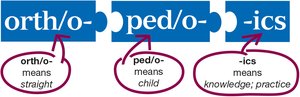
Orth/o: means straight
Ped/o: means child
-ics: means knowledge; practice
Skeletal System Overview
Structure and Function
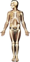
The skeletal system is a widespread, connected body system consisting of 206 bones and other structures. It extends from the top of the head to the tips of the fingers and toes, providing support, protection, and movement.
Key Functions: Support, protection of organs, movement, mineral storage, blood cell production
Major Divisions: Axial skeleton and appendicular skeleton
Anatomy of the Skeleton
Axial Skeleton
The axial skeleton forms the central structure around which other parts move. It includes the head, neck, chest, and spinal bones.
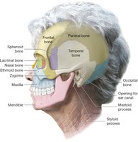
Cranial Bones: Form the cranium and protect the brain
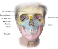
Facial Bones: Support the nose, cheeks, and lips
Ossicles: Three tiny bones in each middle ear (malleus, incus, stapes)
Hyoid Bone: Located in the neck, not connected to other bones
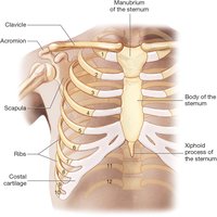
Rib Cage: Sternum and 12 pairs of ribs (true, false, floating)
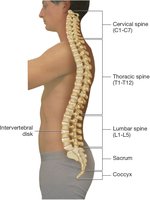
Spine: Five regions: cervical, thoracic, lumbar, sacrum, coccyx
Cranial Bones
Frontal bone: Forehead
Parietal bones: Upper sides and posterior
Occipital bone: Base
Temporal bones: Sides
Sphenoid bone: Central base
Ethmoid bone: Nasal septum
Fontanels
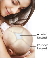
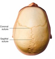
Fontanels are soft spots on an infant's skull where bones have not yet fused. The anterior and posterior fontanels are covered by fibrous connective tissue.
Chest and Rib Cage
Sternum: Manubrium, body, xiphoid process
Ribs: 12 pairs (1-7 true, 8-10 false, 11-12 floating)
Spine
Cervical vertebrae (C1-C7): Neck
Thoracic vertebrae (T1-T12): Thorax
Lumbar vertebrae (L1-L5): Lower back
Sacrum: Five fused vertebrae
Coccyx: Fused vertebrae forming the tailbone
Appendicular Skeleton
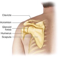
The appendicular skeleton consists of the limbs and their girdles, including the shoulder, arm, hip, and leg bones.
Shoulder
Clavicle (collarbone)
Scapula (shoulder blade)
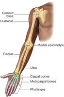
Upper Extremity
Humerus: Upper arm bone
Radius and Ulna: Forearm bones
Carpal bones: Wrist
Metacarpal bones: Hand
Phalanges: Fingers (three per finger, two in thumb)
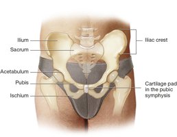
Hip
Ilium: Most superior
Ischium: Most inferior
Pubis: Most anterior
Acetabulum: Hip socket
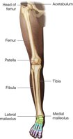
Lower Extremity
Femur: Thigh bone
Tibia: Shin bone
Fibula: Thin bone of lower leg
Patella: Kneecap
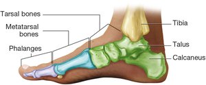
Ankle and Foot
Tarsal bones: Ankle
Metatarsal bones: Midfoot
Phalanges: Toes (three per toe, two in hallux/great toe)
Structure of Bone
Osseous Tissue and Bone Anatomy
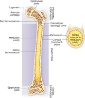
Bone is a type of connective tissue called osseous tissue. The surface is covered by periosteum, a thick, fibrous membrane. Long bones have a shaft (diaphysis) and ends (epiphyses), with cortical (compact) bone and cancellous (spongy) bone.
Medullary cavity: Contains yellow bone marrow
Epiphyseal plates: Sites of bone growth
Red bone marrow: Produces blood cells
Joints, Cartilage, and Ligaments
Types of Joints
Joints (articulations) are formed where two bones meet. They are classified by their movement:
Suture joints: Immovable (cranial and facial bones)
Symphysis joints: Slightly movable (pubis, vertebrae)
Synovial joints: Fully movable; hinge and ball-and-socket types
Joint Structure
Cartilage: Dense collagen fibers, covers bone ends
Meniscus: Cartilage pad in some synovial joints
Ligaments: Strong connective tissue bands
Joint capsule: Contains synovial membrane and fluid
*Additional info: Synovial fluid lubricates and nourishes the joint.*
Physiology of Bone Growth
Bone Cells and Remodeling
Bone growth and maintenance involve several cell types:
Osteocytes: Mature bone cells, monitor mineral content
Osteoclasts: Break down bone tissue
Osteoblasts: Build new bone tissue
Bone is composed of calcium salts and collagen fibers. Calcium is released as needed for muscle function and bone repair.
Bone Remodeling
10% of skeleton is remodeled annually
Osteoclasts dissolve old bone; osteoblasts form new bone (osteogenesis)
In children, bone formation exceeds breakdown; in adults, rates are equal; in older adults, breakdown exceeds formation
Ossification and Growth
Cartilage is replaced by bone at epiphyseal plates
Growth occurs until adulthood
Bone and Cartilage Diseases
Fractures
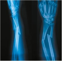
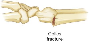
Fractures are breaks in bone, classified by their pattern and severity.
Type | Description |
|---|---|
Colles Fracture | Distal radius fracture, often from a fall |
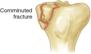
Comminuted Fracture | Bone shattered into multiple pieces |
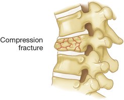
Compression Fracture | Bone is crushed, often in vertebrae |
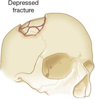
Depressed Fracture | Bone is pressed inward, often in skull |
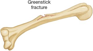
Greenstick Fracture | Partial fracture, common in children |
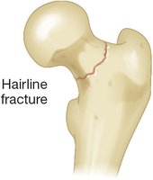
Hairline Fracture | Thin fracture line, minimal displacement |
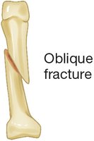
Oblique Fracture | Diagonal break across bone |
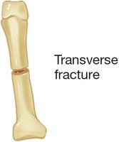
Transverse Fracture | Horizontal break across bone |
Osteoporosis
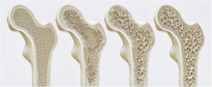
Osteoporosis is a disease characterized by decreased bone density and increased risk of fractures. It is common in older adults, especially postmenopausal women.
Chest and Spine Diseases
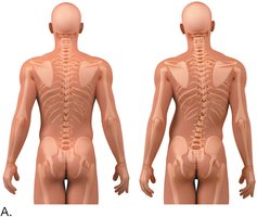
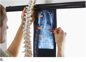
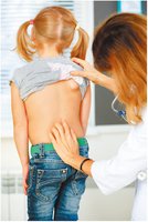
Scoliosis
Scoliosis is a lateral curvature of the spine, which can cause uneven shoulders, hips, and arm lengths. Diagnosis and treatment planning involve physical examination and imaging.
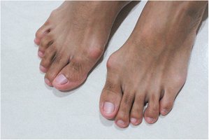
Hallux Valgus and Clubfoot
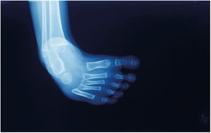
Hallux Valgus: Lateral deviation of the great toe, often with bunion formation
Clubfoot: Congenital deformity with inwardly rotated foot
Joints, Cartilage, and Ligaments Diseases
Osteoarthritis
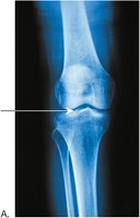
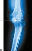
Osteoarthritis is a degenerative joint disease characterized by loss of articular cartilage, narrowing of joint space, and malalignment of bone ends.
Rheumatoid Arthritis
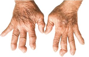
Rheumatoid arthritis is an autoimmune disease causing severe joint deformities, especially in the hands.
Laboratory, Diagnostic, and Radiologic Procedures
Bone Densitometry
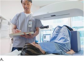
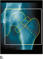
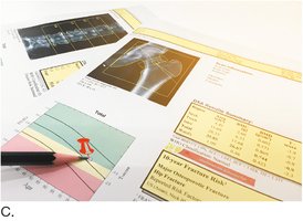
Bone mineral density tests (such as DXA scans) assess bone health and risk of osteoporosis. The hip and spine are common sites for measurement.
Medical Procedures
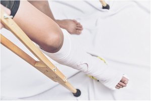
Cast Application and Crutches
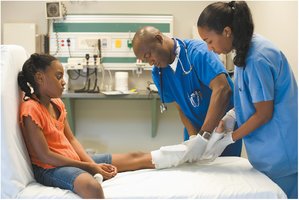
Casts are applied to immobilize fractured bones. Patients may use crutches and require physical therapy after healing.
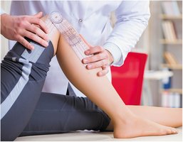
Goniometer
A goniometer is used by physical therapists to measure the range of motion in joints.
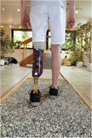
Prosthesis
Prosthetic limbs are custom-built for patients after amputation, allowing for rehabilitation and mobility.
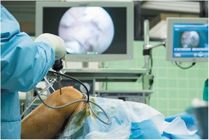
Surgical Procedures
Arthroscopic Surgery
Arthroscopy is a minimally invasive surgical procedure used to examine and treat joint conditions. A fiberoptic scope and instruments are inserted through small portals.
Hip Prostheses
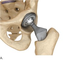
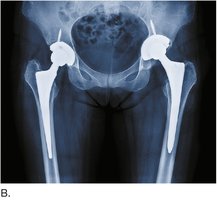
Total hip replacement involves removing the head of the femur and replacing it with a prosthesis. The acetabulum is also replaced with a cup, and the ball is inserted into the cup.
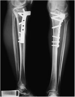
Orthopedic Plate and Screws
Fractures may be stabilized using metal plates and screws.
Appendix: Key Abbreviations and Terms
RUE: Right Upper Extremity
LUE: Left Upper Extremity
RLE: Right Lower Extremity
LLE: Left Lower Extremity
BMI: Body Mass Index
Example Calculation: BMI for Children
For children and teenagers, BMI is calculated as:
*Additional info: Percentiles are used to assess growth and nutrition in pediatric patients.*
