 Indietro
Pulmonology: The Respiratory System – Medical Terminology Study Guide
Guida di studio - Note intelligenti
 Appunti personalizzati basati sui tuoi materiali, ampliati con definizioni chiave, esempi e contesto.
Pulmonology and the Respiratory System
Introduction to Pulmonology
Pulmonology is the medical specialty focused on the anatomy, physiology, diseases, and treatments of the respiratory system. Pulmonologists utilize diagnostic tests, medical and surgical procedures, and pharmacological interventions to manage respiratory conditions.
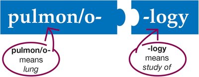
Pulmon/o-: means lung
-logy: means study of
Respiratory system: includes the lungs and associated structures forming the pathway for air flow.
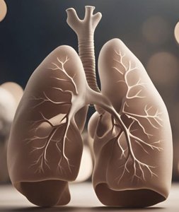
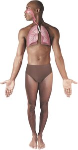
Anatomy of the Respiratory System
Upper Respiratory System
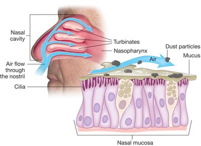
The upper respiratory tract consists of the nose, nasal cavity, and pharynx. These structures filter, warm, and moisten incoming air, protecting the lower respiratory tract.
Nose and Nasal Cavity: Entry point for air; divided by the septum.
Turbinates (Nasal Conchae): Bony projections that slow air flow, aiding warming and moistening.
Nasal Mucosa: Mucous membrane lining the cavity and turbinates; humidifies air and traps particles.
Cilia and Hair: Trap and clear debris from inhaled air.
Pharynx
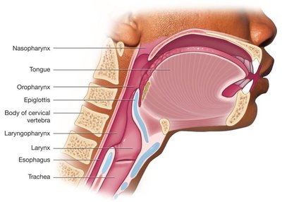
The pharynx is a muscular tube serving as a common passage for air and food. It is divided into three regions: nasopharynx, oropharynx, and laryngopharynx.
Nasopharynx: Uppermost part, behind the nasal cavity.
Oropharynx: Middle part, behind the oral cavity.
Laryngopharynx: Lowest part, leading to the larynx and esophagus.
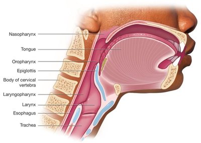
Larynx (Voice Box)
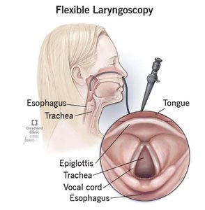
The larynx remains open during breathing and contains the vocal cords, which vibrate to produce sound. During swallowing, the larynx moves upward to meet the epiglottis, preventing food from entering the airway.
Vocal cords: Bands of smooth muscle responsible for sound production.
Epiglottis: Flap that covers the larynx during swallowing.
Lower Respiratory System
Trachea, Bronchi, and Bronchioles
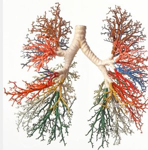
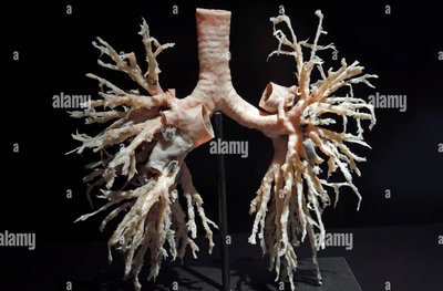
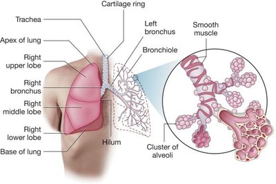
The trachea (windpipe) is a rigid tube supported by C-shaped cartilage rings, dividing into right and left bronchi. Bronchi further branch into bronchioles, which lack cartilage and have smooth muscle walls. The bronchial tree is lined with cilia to clear debris.
Trachea: Main airway, divides into bronchi.
Bronchi: Right and left branches entering the lungs.
Bronchioles: Smallest airways, ending in alveoli.
Lungs and Alveoli
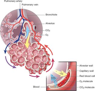
The lungs are spongy, air-filled organs. The right lung has three lobes; the left lung has two. Bronchioles terminate in alveoli, which are hollow spheres where gas exchange occurs. Alveoli secrete surfactant to prevent collapse.
Right lung: Upper, middle, and lower lobes.
Left lung: Upper and lower lobes.
Alveoli: Site of oxygen and carbon dioxide exchange with capillaries.
Pulmonary parenchyma: Functional tissue formed by alveoli.
Thorax and Pleura
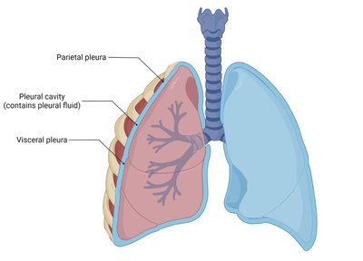
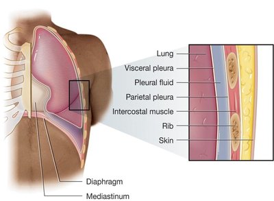
The thoracic cavity houses the lungs, heart, and other structures. Each lung is contained within a pleural cavity, lined by a double-layered pleura that secretes fluid for smooth movement during breathing.
Rib cage: Sternum, ribs, and spine protect the lungs.
Mediastinum: Central area containing the heart, trachea, and esophagus.
Diaphragm: Muscle forming the inferior border, essential for breathing.
Pleura: Visceral (lung surface) and parietal (thoracic wall) layers.
Physiology of Respiration
Breathing Mechanics
Breathing is regulated involuntarily by control centers in the brain via the phrenic nerve. Inhalation involves contraction of the diaphragm and intercostal muscles, expanding the thoracic cavity and creating negative pressure. Exhalation is the relaxation of these muscles, reducing cavity size and expelling air.
Inhalation (Inspiration): Diaphragm contracts, thoracic cavity enlarges, air flows in.
Exhalation (Expiration): Diaphragm relaxes, thoracic cavity shrinks, air flows out.
Forceful exhalation: Abdominal and intercostal muscles contract to expel air rapidly.

Processes of Respiration
Respiration involves five key processes:
Ventilation: Movement of air in and out of the lungs.
External respiration: Gas exchange between alveoli and blood.
Gas transport: Movement of gases via blood.
Internal respiration: Oxygen transfer from blood to cells.
Cellular respiration: Oxygen used for metabolism; carbon dioxide produced as waste.
Diseases and Disorders of the Respiratory System
Upper Respiratory Diseases
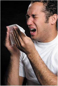
Common conditions include upper respiratory infections (URIs), which are often viral or bacterial and spread easily through contact or airborne droplets.
URI: Head cold; antibiotics only effective for bacterial causes.
Lower Respiratory Diseases
Diseases affecting the trachea, bronchi, bronchioles, and lungs include asthma, bronchiectasis, bronchitis, and chronic obstructive pulmonary disease (COPD).
Asthma: Hyperreactivity causing inflammation, mucus, and bronchospasm.
Bronchiectasis: Permanent dilation and loss of elasticity in bronchioles.
Bronchitis: Infection or inflammation of bronchi; acute (infection) or chronic (smoking/pollution).
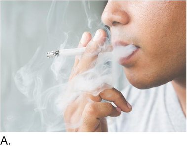
COPD: Chronic bronchitis and emphysema; characterized by inflammation, cough, and barrel chest.
Lung Diseases
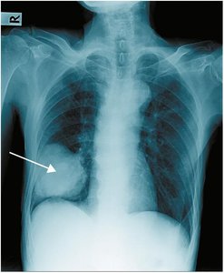
Lung diseases include pneumonia, pulmonary edema, pulmonary embolism, tuberculosis, and occupational lung diseases.
Pneumonia: Infection filling lung passages with fluid and cells; can be bacterial, viral, or fungal.
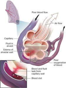
Pulmonary edema: Fluid accumulation in alveoli, often due to heart failure.
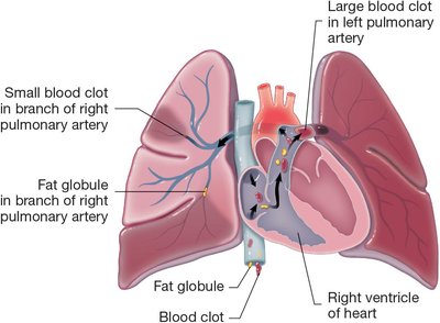
Pulmonary embolism: Blockage of pulmonary artery by embolus, reducing oxygenation.
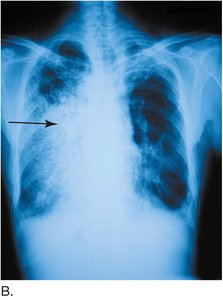
Tuberculosis (TB): Caused by Mycobacterium tuberculosis; forms nodules and is drug-resistant.
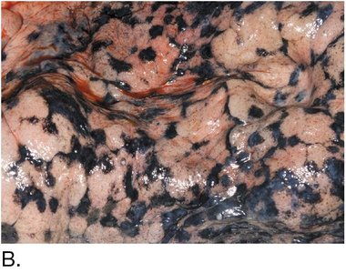
Occupational lung diseases: Caused by inhaled particles (e.g., anthracosis, asbestosis).
Thoracic Diseases
Conditions affecting the thoracic cavity include hemothorax, pleural effusion, pleurisy, and pneumothorax.
Hemothorax: Blood in the thoracic cavity, usually from trauma.
Pleural effusion: Excess fluid in pleural space.
Pleurisy: Inflammation of pleura causing friction rub.
Pneumothorax: Air in pleural space causing lung collapse.
Respiratory Disorders
Disorders of breathing include apnea, bradypnea, cough, dyspnea, orthopnea, and tachypnea.
Apnea: Absence of breathing.
Bradypnea: Slow breathing rate.
Cough: Protective mechanism to expel substances.
Dyspnea: Difficult or labored breathing.
Orthopnea: Difficulty breathing when supine.
Tachypnea: Rapid breathing rate.
Oxygen and Carbon Dioxide Disorders
Imbalances in oxygen and carbon dioxide levels include anoxia, asphyxia, cyanosis, hypercapnia, and hypoxemia.
Anoxia: Complete lack of oxygen.
Asphyxia: High CO2, low O2 in blood.
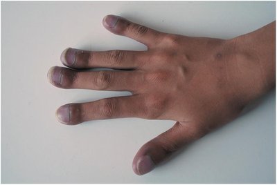
Cyanosis: Bluish skin due to low oxygen.
Hypercapnia: High CO2 in blood.
Hypoxemia: Low O2 in blood.
Laboratory Tests and Diagnostic Procedures
Common Tests
Arterial blood gases (ABGs): Measures O2 and CO2 in arterial blood.
Carboxyhemoglobin: Measures CO in blood.
Oximetry: Measures oxygen saturation noninvasively.
Pulmonary function test (PFT): Measures lung volumes and capacities.
Sleep study: Assesses sleep apnea.
Sputum culture & sensitivity (C&S): Identifies causative bacteria and antibiotic sensitivity.
Tuberculosis test: Detects exposure to TB.
Radiologic and Nuclear Medicine Procedures
Chest radiography (CXR): X-ray imaging of lungs.
Computerized axial tomography (CAT/CT): Cross-sectional imaging.
Lung scan: Identifies areas of poor gas exchange.
Magnetic resonance imaging (MRI): Detailed tissue imaging.
Medical and Surgical Procedures
Medical Procedures
Auscultation and percussion (A&P): Listening to breath sounds and tapping for dullness.
Cardiopulmonary resuscitation (CPR): Emergency procedure for cardiac and respiratory arrest.
Chest percussion therapy: Loosens mucus for expectoration.
Continuous positive airway pressure (CPAP): Treats sleep apnea.
Endotracheal intubation: Establishes airway for oxygen delivery.
Incentive spirometry: Encourages deep breathing post-surgery.
Nebulizer treatment: Delivers medication as mist.
Oxygen therapy: Provides supplemental oxygen.
Surgical Procedures
Bronchoscopy: Examines trachea and bronchi.
Chest tube insertion: Removes air, fluid, pus, or blood.
Lung resection: Removes part or all of a lung.
Thoracentesis: Removes pleural fluid.
Thoracotomy: Incision into thoracic cavity.
Tracheostomy: Creates airway access.
Drugs Used in Respiratory Medicine
Antibiotic drugs: Treat bacterial infections.
Antitubercular drugs: Treat tuberculosis.
Antitussive drugs: Suppress cough.
Antiviral drugs: Treat viral infections.
Bronchodilator drugs: Relax smooth muscle, widen airways.
Corticosteroid drugs: Reduce inflammation.
Expectorants: Thin mucus for easier expulsion.
Leukotriene receptor blockers: Reduce inflammation in asthma.
Smoking cessation drugs: Aid in quitting smoking.
Abbreviation Summary
Abbreviation | Definition |
|---|---|
A&P | auscultation and percussion |
ABG | arterial blood gases |
CXR | chest x-ray |
COPD | chronic obstructive pulmonary disease |
CPAP | continuous positive airway pressure |
CT | computerized axial tomography |
O2 | oxygen |
PFT | pulmonary function test |
TB | tuberculosis |
URI | upper respiratory infection |
