 Indietro
Skeletal System: Structure, Function, and Clinical Relevance
Guida di studio - Note intelligenti
 Appunti personalizzati basati sui tuoi materiali, ampliati con definizioni chiave, esempi e contesto.
The Skeletal System
Overview and Functions
The skeletal system is composed of bones and connective tissues such as cartilage and ligaments. It provides the framework for the body, protects vital organs, enables movement, stores minerals, and houses sites for blood cell production.
Support and Protection: Bones support body structures and protect internal organs (e.g., skull protects the brain, rib cage protects the heart and lungs).
Movement: Bones act as levers for muscles, facilitating movement.
Storage: Bones store calcium ions (Ca2+), phosphate (PO43-), and lipids (in yellow marrow).
Blood Cell Production (Hematopoiesis): Occurs in red bone marrow.
Axial vs. Appendicular Skeleton
Classification of Bones
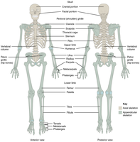
The human skeleton is divided into two main parts: the axial skeleton and the appendicular skeleton.
Axial Skeleton: Consists of the skull, vertebral column, and rib cage. It forms the central axis of the body.
Appendicular Skeleton: Includes the bones of the limbs and girdles (shoulder and pelvic girdles) that attach them to the axial skeleton.
Bone Tissue Types: Compact vs. Spongy Bone
Structural Differences and Locations
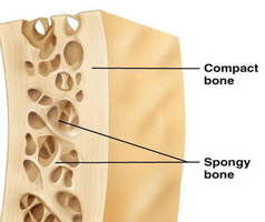
Bones are composed of two main types of tissue: compact bone and spongy bone, each with distinct characteristics and functions.
Compact Bone: Dense and strong, found on the external surfaces of bones. Organized into osteons, providing rigidity and support.
Spongy Bone: Porous and lighter, located internally. Composed of trabeculae, with spaces filled by red or yellow bone marrow.
Structure of a Long Bone
Major Anatomical Features
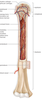
Long bones, such as the femur and humerus, have specialized structures that support their function and growth.
Epiphysis: The expanded ends of the bone, covered with articular cartilage for joint movement.
Diaphysis: The shaft of the bone, containing the medullary cavity.
Epiphyseal Plate/Line: Growth plate made of cartilage in children; becomes a bone line in adults.
Medullary Cavity: Central cavity containing yellow marrow (fat storage).
Periosteum: Fibrous membrane covering the bone, containing nerves, blood vessels, and bone stem cells.
Red Bone Marrow Locations
Sites of Hematopoiesis
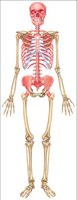
In adults, red bone marrow is primarily found in the spongy bone of the axial skeleton, os coxae, and the heads of the femur and humerus.
Microscopic Structure of Bone
Osteons and Cellular Components
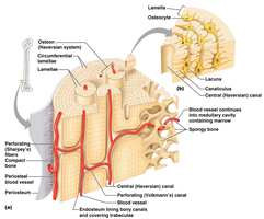
Bone tissue is organized into microscopic units called osteons, which provide strength and facilitate nutrient delivery.
Osteons: Cylindrical structures running parallel to the bone's long axis.
Concentric Lamellae: Rings of bone matrix around the central canal.
Central Canal: Contains blood vessels and nerves.
Osteocytes: Mature bone cells residing in lacunae.
Canaliculi: Tiny channels allowing nutrient and waste exchange between osteocytes and the central canal.
Bone Cells and Extracellular Matrix
Cell Types and Matrix Composition
Bone tissue contains specialized cells and a matrix that provides both strength and flexibility.
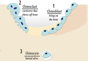
Osteoblasts: Cells that synthesize and secrete bone matrix (bone formation).
Osteocytes: Mature osteoblasts that maintain bone tissue.
Osteoclasts: Large cells that resorb (break down) bone matrix, releasing calcium into the blood.
Extracellular Matrix: Composed of inorganic calcium phosphate salts (resist compression) and organic collagen fibers (provide tensile strength).
Bone Remodeling and Health
Diet, Exercise, and Bone Adaptation
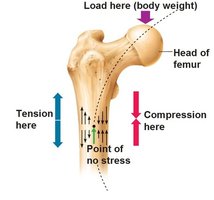
Bone is a dynamic tissue that remodels in response to stress and nutritional factors, following Wolff’s Law.
Wolff’s Law: Bone grows and remodels in response to the forces placed upon it.
Exercise: Increases bone density and strength.
Calcium and Vitamin D: Essential for bone matrix formation and maintenance.
Protein: Necessary for collagen synthesis.
Osteoporosis
Pathophysiology and Risk Factors
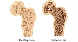
Osteoporosis is a disorder characterized by decreased bone mass and increased fracture risk, especially in postmenopausal women due to hormonal changes.
Healthy Bone vs. Osteoporotic Bone: Osteoporotic bone has larger spaces and thinner trabeculae, making it weaker.
Prevention: Adequate calcium, vitamin D, and weight-bearing exercise are important for prevention.
Bone Fracture Repair
Stages of Healing
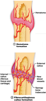
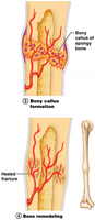
Bone repair occurs in four main stages following a fracture:
Hematoma Formation: Blood vessels rupture, forming a hematoma at the fracture site.
Cartilaginous (Fibrocartilaginous) Callus Formation: Cartilage forms to bridge the broken bone ends.
Bony Callus Formation: Osteoblasts replace cartilage with spongy bone.
Bone Remodeling: Osteoclasts and osteoblasts reshape the bone to its original form.
Joints (Articulations)
Classification and Structure
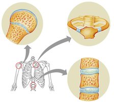
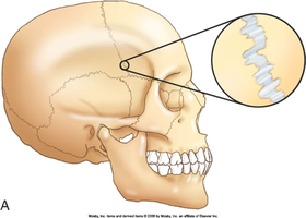
Joints are sites where two bones meet, classified by their structure and the amount of movement they allow.
Fibrous Joints: Bones joined by fibrous tissue; immovable (e.g., sutures of the skull).
Cartilaginous Joints: Bones joined by cartilage; slightly movable (e.g., intervertebral discs, pubic symphysis).
Synovial Joints: Bones separated by a fluid-filled cavity; freely movable (e.g., knee, shoulder).
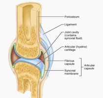
Structure of Synovial Joints
Key Components
Synovial joints are the most common and movable type of joint in the body, characterized by several specialized structures:
Fibrous Capsule: Encloses the joint, providing stability.
Articular Cartilage: Covers bone ends, reducing friction.
Synovial Membrane: Secretes synovial fluid for lubrication.
Joint Cavity: Space filled with synovial fluid.
Ligaments: Connect bone to bone, stabilizing the joint.
Tendons: Connect muscle to bone, aiding movement.
Major Synovial Joint Movements
Types of Movements
Synovial joints allow for a variety of movements, including:
Flexion: Decreases the angle between bones (e.g., bending the elbow).
Extension: Increases the angle between bones (e.g., straightening the knee).
Adduction: Moves a limb toward the midline of the body.
Abduction: Moves a limb away from the midline.
Elevation/Depression: Moves a body part superiorly (up) or inferiorly (down), such as the shoulders or mandible.
Sprains, Dislocations, and Arthritis
Common Joint Disorders
Sprain: Stretching or tearing of ligaments reinforcing a joint.
Dislocation: Bones forced out of their normal position, often accompanied by ligament injury.
Osteoarthritis: Degenerative joint disease caused by wear and tear of articular cartilage.
Rheumatoid Arthritis: Autoimmune disorder affecting the synovial membrane, leading to inflammation and joint damage.
Summary Table: Bone Cell Types and Functions
Cell Type | Function |
|---|---|
Osteoblast | Bone formation (secretes bone matrix) |
Osteocyte | Maintains bone tissue |
Osteoclast | Bone resorption (breaks down bone matrix) |
