 Indietro
Study Guide: Medical Terminology – Muscular System (Orthopedics)
Guida di studio - Note intelligenti
 Appunti personalizzati basati sui tuoi materiali, ampliati con definizioni chiave, esempi e contesto.
Orthopedics and the Muscular System
Overview of Orthopedics
Orthopedics is the medical specialty focused on the study and treatment of the muscular and skeletal systems. This field utilizes laboratory and diagnostic tests, medical and surgical procedures, and pharmacological interventions to address diseases affecting these systems. In this chapter, the emphasis is placed on the muscular system.
Anatomy of the Muscular System
Components of the Muscular System
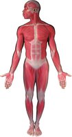
The muscular system consists of muscles, tendons, and associated connective tissues throughout the body. Its primary functions are to maintain body position and facilitate movement of the skeletal framework. Due to its close association with bones, it is often referred to as the musculoskeletal system.
Muscles: Contractile tissues responsible for movement.
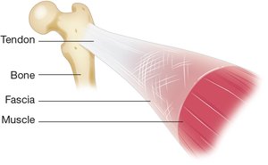
Tendons: Fibrous bands connecting muscles to bones.
Fascia: Thin connective tissue wrapping each muscle and merging into tendons.
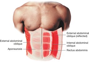
Aponeurosis: Broad, flat sheet of connective tissue attaching flat muscles to bones or deeper muscles.
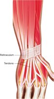
Retinaculum: Translucent band holding down tendons at the wrist and ankle.
Bursa: Fluid-filled sac reducing friction where tendons rub against bones.
Types of Muscle Tissue
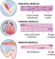
There are three types of muscle tissue in the human body, each with distinct structure and function:
Skeletal Muscle: Voluntary muscles responsible for body movement; characterized by striations and multiple nuclei.
Cardiac Muscle: Involuntary muscle forming the heart; has less prominent striations and typically a single nucleus.
Smooth Muscle: Involuntary muscle forming a thin layer around organs; lacks striations and has a single nucleus.
Only skeletal muscle is considered part of the muscular system for movement.
Muscle Origins and Insertions
Muscles attach to bones at two primary sites:
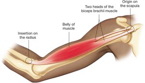
Origin: The site where a tendon attaches to a stationary or nearly stationary bone.
Insertion: The site where a tendon attaches to the bone that moves during muscle contraction.
Belly: The thickest part of the muscle, containing the greatest mass.
Muscle Naming Conventions
Latin Roots and Word Parts
Muscle names are derived from Latin and often describe their location, shape, size, or action. Understanding these roots aids in deciphering medical terminology.
Biceps brachii: 'Biceps' means two heads; 'brachii' means of the arm.
Brachioradialis: 'Brachi/o-' means arm; 'radi/o-' means radius (forearm bone); '-alis' means pertaining to.
Extensor digitorum: 'Extens/o-' means straightening; '-or' means one who does.
Flexor hallucis brevis: 'Flex/o-' means bending; 'hallucis' refers to the big toe; 'brevis' means short.
Gluteus maximus: 'Gluteus' means buttocks; 'maximus' means large.
Rectus abdominis: 'Rectus' means straight; 'abdominis' means of the abdomen.
Temporalis: Pertains to the side of the head.
Triceps brachii: 'Tri-' means three; '-ceps' from 'caput' (head).
Major Muscles of the Body
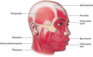
Muscles of the Head and Neck
These muscles facilitate facial expressions, chewing, and head movement.
Temporalis
Masseter
Sternocleidomastoid
Platysma
Frontalis
Orbicularis oculi
Buccinator
Orbicularis oris
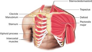
Muscles of the Shoulders and Chest
These muscles are involved in arm movement, shoulder rotation, and breathing.
Sternocleidomastoid
Trapezius
Deltoid
Pectoralis major
Intercostal muscles
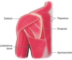
Muscles of the Shoulders and Back
These muscles support posture and facilitate movements such as shrugging and trunk rotation.
Deltoid
Trapezius
Latissimus dorsi
Scapula
Aponeurosis
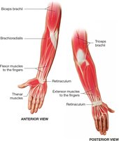
Muscles of the Upper Extremity
These muscles allow for complex movements of the arms, hands, and fingers.
Biceps brachii
Brachioradialis
Flexor muscles
Extensor muscles
Thenar muscles
Triceps brachii
Retinaculum
Muscles of the Forearm and Retinaculum
The retinaculum holds tendons in place, allowing for efficient finger movement.
Extensor muscles
Retinaculum
Muscles of the Abdomen
These muscles are essential for trunk movement, posture, and abdominal compression.
External abdominal oblique
Internal abdominal oblique
Rectus abdominis
Aponeurosis
Muscles of the Lower Extremity
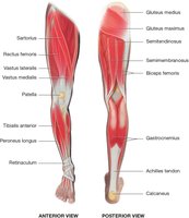
These muscles facilitate leg movement, walking, and balance.
Sartorius
Rectus femoris
Vastus lateralis
Vastus medialis
Patella
Tibialis anterior
Peroneus longus
Gluteus medius
Gluteus maximus
Semitendinosus
Semimembranosus
Biceps femoris
Gastrocnemius
Achilles tendon
Calcaneus
Muscle Strength and Size
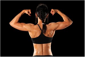
Muscle strength and size are influenced by genetics, nutrition, and physical activity. Highly developed muscles, such as those seen in bodybuilders, demonstrate the adaptability of skeletal muscle tissue.
Muscle Structure and Function
Microscopic Anatomy of Muscle
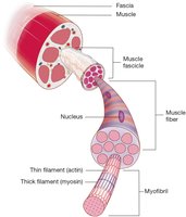
Muscles are composed of fascicles, which contain bundles of muscle fibers (cells). Each fiber contains myofibrils made up of actin (thin) and myosin (thick) filaments, which are essential for contraction.
Fascicle: Bundle of muscle fibers wrapped in fascia.
Muscle fiber: Long cell with multiple nuclei.
Myofibril: Contains actin and myosin filaments.
Neuromuscular Junction and Muscle Contraction
Each muscle fiber is connected to a nerve cell at the neuromuscular junction. The nerve releases acetylcholine, which increases the fiber's permeability and triggers calcium ion release. Calcium enables actin to slide between myosin, shortening the muscle and producing contraction.
Acetylcholine: Neurotransmitter initiating muscle contraction.
Calcium ions: Essential for actin-myosin interaction.
Muscle Tone
Muscles maintain a mild, partial contraction (muscle tone) due to continuous nerve impulses. This keeps muscles firm and ready for action, and is the only skeletal muscle activity not under conscious control.
Types of Muscle Movement
Movement Terminology
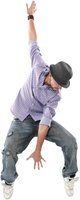
Muscle movements are described using specific terms:
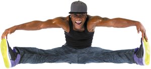
Extension: Straightening a limb.
Abduction: Moving a limb away from the midline.
Adduction: Moving a limb toward the midline.
Flexion: Bending a limb.
Pronation: Rotating the hand or foot downward.
Dorsiflexion: Raising the foot upward.
Plantar flexion: Pointing the foot downward.
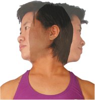
Rotation: Turning a body part around its axis.
Growth and Development Milestones
Muscle Coordination in Infancy
Developmental milestones, such as lifting the head and supporting the upper body, require coordinated muscle activity and strength in the neck, shoulders, and arms.

Diseases of the Muscles and Related Structures
Muscle Atrophy
Muscle atrophy is the wasting or loss of muscle tissue, often due to malnutrition, inactivity, or disease. It results in weakness and decreased muscle mass.

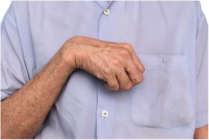
Muscle Contracture
Contracture is the permanent tightening of muscles, tendons, or ligaments, restricting movement. It commonly occurs in patients with limited mobility.
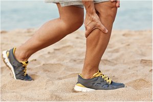
Muscle Strain
Muscle strain is an injury caused by overstretching or tearing muscle fibers, often due to overexertion.
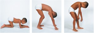
Muscular Dystrophy
Muscular dystrophy is a group of genetic diseases characterized by progressive muscle weakness and degeneration. It often affects the muscles of the legs and arms.
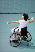
Tennis Elbow
Tennis elbow is an overuse injury causing inflammation of the tendons at the elbow, commonly affecting athletes.
Diseases of the Bursa, Fascia, or Tendon
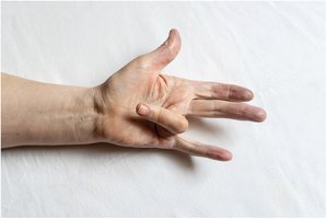
Dupuytren Contracture
Dupuytren contracture is a progressive disease affecting the fingers, causing them to bend toward the palm and limiting extension.
Medical Epicondylitis (Pitcher's Elbow)
Pitcher's elbow is an overuse injury caused by repetitive flexion and gripping, leading to inflammation of the tendons at the elbow.

Medical Procedures and Tests
Braces
Braces are used to provide support and stability to injured or weak muscles and joints, aiding in rehabilitation.

Deep Tendon Reflex
Deep tendon reflex testing assesses the integrity of the neuromuscular system by eliciting involuntary muscle contractions with a percussion hammer.

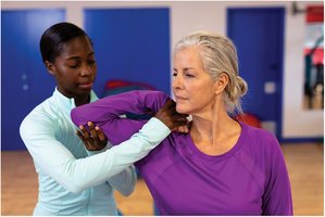
Physical Therapy
Physical therapy is used to decrease muscle stiffness, improve mobility, and restore function after injury or surgery.
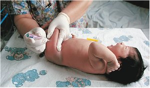
Intramuscular Injection
Intramuscular injections are administered into large muscles, such as the thigh, for rapid absorption of medications, including immunizations in infants.
Summary Table: Types of Muscle Tissue
Type | Location | Control | Appearance | Function |
|---|---|---|---|---|
Skeletal | Attached to bones | Voluntary | Striated, multiple nuclei | Movement of body |
Cardiac | Heart | Involuntary | Striated, single nucleus | Pumping blood |
Smooth | Walls of organs | Involuntary | No striations, single nucleus | Movement of substances |
Key Formula: Muscle Contraction
The sliding filament theory describes muscle contraction:
Calcium ions and acetylcholine are essential for this process.
Additional info: Academic context was added to clarify muscle structure, naming conventions, and disease mechanisms, ensuring completeness and self-contained study notes for exam preparation.
