 Indietro
Acellular Agents: Viruses and Prions – Comprehensive Study Notes
Guida di studio - Note intelligenti
 Appunti personalizzati basati sui tuoi materiali, ampliati con definizioni chiave, esempi e contesto.
Acellular Agents: Viruses and Prions
Characteristics of Viruses
Viruses are unique infectious agents that can infect every type of cell, including bacteria, plants, animals, and fungi. They are not considered living organisms because they cannot exist independently from their host cells and lack essential cellular machinery.
Obligate intracellular parasites: Viruses must invade a host cell to multiply, using the host's genetic and metabolic machinery.
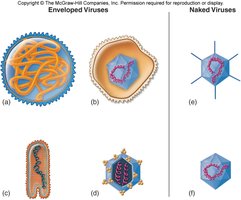
Structure: Some viruses are enveloped, while others are naked. Many have spikes for attachment.
No ribosomes or ATP-generating mechanisms: Viruses rely entirely on the host cell for protein synthesis and energy.
Protein coat: All viruses possess a protein coat (capsid) that protects their genetic material.
Infectious particles: Viruses are referred to as infectious particles rather than cells.
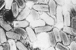
Additional info: Viruses are much smaller than most cells and can only be visualized with electron microscopy.
Visualizing Viruses
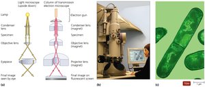
Due to their small size, viruses require specialized techniques for visualization.
Electron microscopy: Most viruses are only visible with electron microscopes.
Staining: Negative staining outlines the shape, while positive staining reveals internal details.
Host Range
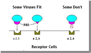
The host range of a virus is determined by specific attachment sites and cellular factors. Viruses may be generalists or specialists.
Generalists: Infect many kinds of cells in various hosts.
Specific: Infect only particular host cells.
Classification and Naming of Viruses
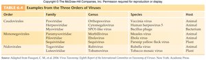
Viruses are classified based on structure, chemical composition, and genetic similarities. The International Committee on the Taxonomy of Viruses (ICTV) organizes viruses into orders, families, genera, and species.
Orders: 3 main orders
Families: 63 families (suffix: -viridae)
Genera: 263 genera (suffix: -virus)
Order | Family | Genus | Species | Host |
|---|---|---|---|---|
Caudovirales | Podoviridae | Orthopoxvirus | Vaccinia virus | Animal |
Mononegavirales | Paramyxoviridae | Rubulavirus | Mumps virus | Animal |
Nidovirales | Coronaviridae | Coronavirus | Avian infectious bronchitis virus | Plant |
Additional info: Table shows examples from three orders of viruses. | ||||
Viral Components
Viruses contain only the parts necessary to infect and control a host cell.
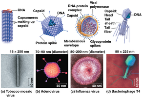
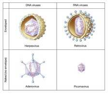
External coating: Capsid, spikes, and envelope (in some viruses).
Core: DNA or RNA.
Nucleocapsid: The capsid and nucleic acid together.
Naked virus: Virus without an envelope.
Functions and Shapes of the Viral Capsid
The capsid protects viral nucleic acids, aids in host cell entry, and stimulates the immune system.
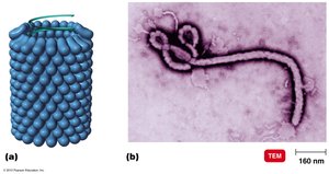
Helical: Rod-shaped, with nucleic acid inside a hollow cylinder.
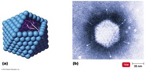
Polyhedral: Many-sided, often icosahedral.
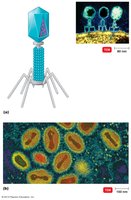
Complex: Not strictly helical or polyhedral; includes bacteriophages.
Helical Virus Example
Ebola virus is a classic example of a helical virus.
Polyhedral Virus Example
Mastadenovirus is a typical polyhedral virus.
Complex Virus Example
Bacteriophages and orthopoxviruses are examples of complex viruses.
Nucleic Acids at the Core of a Virus
Viruses have a small number of genes, just enough to invade host cells and redirect their activity. There are two main types:
DNA viruses
RNA viruses
Types of DNA Viruses
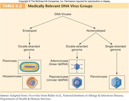
DNA viruses can be enveloped or non-enveloped, and may have double- or single-stranded genomes.
Types of RNA Viruses
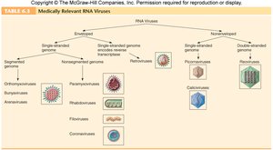
Most RNA viruses are single-stranded. Positive-sense RNA genomes are ready for translation, while negative-sense RNA genomes must be converted before translation. Some RNA viruses are segmented.
Modes of Viral Multiplication
Viral multiplication requires a host cell. Bacteriophages and animal viruses have distinct replication cycles.
Bacteriophages: Lytic and lysogenic cycles.
Animal viruses: Lysis and budding.
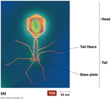
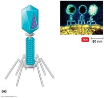
Bacteriophage Structure
Bacteriophages have an icosahedral capsid head containing DNA, a central tube (sheath), neck, base plate, tail pins, and fibers.
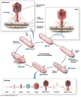
Lytic Cycle of Bacteriophages
The lytic cycle results in the destruction of the host cell and release of new phages.
Attachment: Phage attaches to host cell.
Entry: Phage injects DNA.
Synthesis: Host cell produces viral components.
Assembly: New phages are assembled.
Release: Host cell lyses, releasing phages.
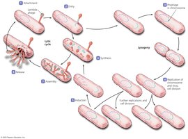
Lysogenic Cycle of Bacteriophages
In the lysogenic cycle, viral DNA integrates into the host genome and is replicated along with it.
Multiplication of Animal Viruses
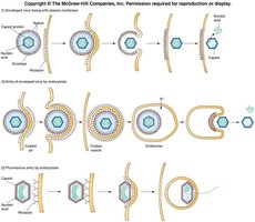
Animal viruses replicate through a series of steps: attachment, penetration, uncoating, biosynthesis, maturation, and release.
Attachment: Virus binds to cell membrane.
Penetration: Entry by endocytosis, direct penetration, or fusion.
Uncoating: Viral or host enzymes remove capsid.
Biosynthesis: Production of viral nucleic acids and proteins.
Maturation/Assembly: Assembly of viral components.
Release: By budding (enveloped viruses) or rupture.
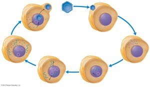
Replication of a DNA-Containing Animal Virus
Papovavirus is a typical DNA-containing virus that attacks animal cells. The replication process involves attachment, entry, uncoating, biosynthesis, maturation, and release.
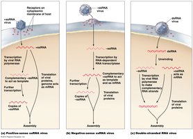
Life Cycle of Animal Viruses (Synthesis – RNA Viruses)
RNA viruses synthesize their genomes and proteins differently depending on whether they are positive-sense, negative-sense, or double-stranded.
Life Cycle of Animal Viruses (Exit from Host)
Enveloped viruses exit by budding, while non-enveloped viruses typically exit by cell rupture.
Latency of Animal Viruses
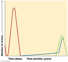
Some viruses can lie dormant within a cell, known as latency. Latent viruses (proviruses) include herpesviruses and chickenpox. HIV can also integrate into the host genome.
Latent infection: Virus remains inactive for extended periods.
Persistent infection: Virus is continuously released.
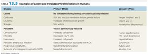
Disease | Primary Effect | Causative Virus |
|---|---|---|
Cold sores | No symptoms during latency | Herpes simplex 1 |
Leukemia | Virus continuously released | HTLV-1 |
Measles | Mental deterioration | Measles virus |
Additional info: Table shows examples of latent and persistent viral infections in humans. | ||
Viruses and Cancer
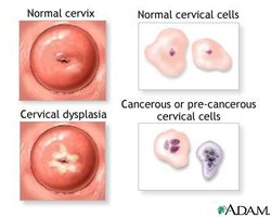
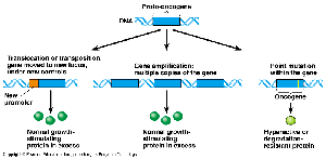
Viruses are implicated in 20-25% of human cancers. Oncogenic viruses can activate oncogenes, leading to uncontrolled cell division (neoplasia).
Examples: Hodgkin’s disease, cervical cancer, Kaposi’s sarcoma.
Transformed cells: Increased growth, loss of contact inhibition, tumor-specific antigens.
Integration: Viral genetic material integrates into host DNA.
Culturing Viruses
Viruses can be cultured using various methods, each with advantages and disadvantages.
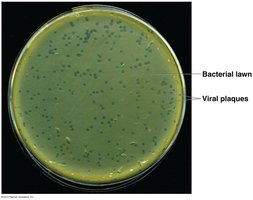
Bacteriophages: Cultured on bacterial lawns; plaques indicate viral activity.
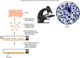
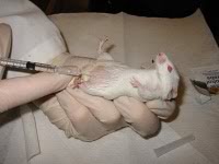
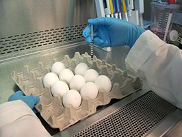
Animal viruses: Tissue culture, live animals, and embryonated eggs.
Applications: Vaccine production (e.g., influenza).
Virus Identification
Viruses are identified using serological tests (detecting antibodies), nucleic acid analysis (RFLPs, PCR), and other molecular techniques.
Serological tests: Neutralization, hemagglutination, Western blot.
Nucleic acid tests: Restriction fragment length polymorphism (RFLP), polymerase chain reaction (PCR).
Prions
Prions are infectious proteins that cause neurodegenerative diseases. Unlike viruses, prions lack nucleic acids and propagate by inducing misfolding of normal proteins.
Diseases: Creutzfeldt-Jakob disease, mad cow disease.
Mechanism: Prion proteins convert normal proteins into abnormal forms.
Key Points to Know
Understand the composition and life cycle of enveloped and non-enveloped animal viruses and bacteriophages.
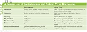
Compare and contrast bacteriophage and animal virus life cycles.
Know viral shapes and capsid morphology.
Understand virus classification and taxonomy.
Explain methods for culturing viruses and their pros/cons.
Discuss prion infectivity.
