 Indietro
Acellular Infectious Agents: Viruses, Prions, and Viroids – Structure, Genomes, and Life Cycles
Guida di studio - Note intelligenti
 Appunti personalizzati basati sui tuoi materiali, ampliati con definizioni chiave, esempi e contesto.
Acellular Infectious Agents
Types and Basic Properties
Acellular infectious agents are pathogens that lack cellular structure and depend entirely on living cells for replication. There are three main types: viruses, prions, and viroids. Each has distinct structural and functional characteristics.
Viruses: Infectious particles composed of a nucleic acid genome (DNA or RNA) wrapped in a protein coat (capsid); some have a lipid envelope.
Prions: Infectious proteins lacking nucleic acids; cause neurodegenerative diseases.
Viroids: Infectious nucleic acid genomes (usually RNA) without a protein coat; primarily infect plants.
All acellular agents require a host cell for replication and cannot reproduce independently.
They can infect a wide range of organisms, including bacteria, fungi, plants, and animals.
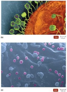
Viral Structure
Virion Components and Shapes
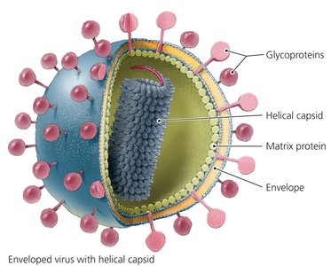
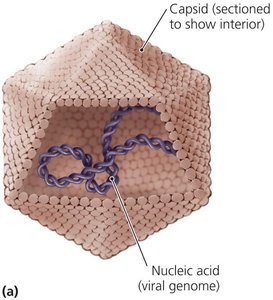
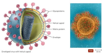
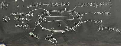
The extracellular form of a virus is called a virion. Virions must contain a nucleic acid genome and a protein coat called a capsid. The combination of genome and capsid is termed the nucleocapsid. Some viruses possess an additional lipid envelope derived from the host cell membrane.
Capsid: Made of protein subunits called capsomeres; protects and contains the viral genome.
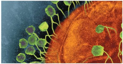
Capsid Shapes: Common shapes include helical (rod-shaped), polyhedral (often icosahedral), and complex (e.g., bacteriophage structure).
Envelope: Lipid bilayer surrounding some nucleocapsids; contains embedded proteins from both host and virus.
Matrix proteins: Located between the nucleocapsid and envelope; contribute to virion stability.
Viral Genomes
Types and Classification
Viral genomes are much smaller than those of living cells and encode fewer proteins. The genome type is a key determinant of viral replication mechanisms and classification.
Can be DNA or RNA.
Can be double-stranded (ds) or single-stranded (ss).
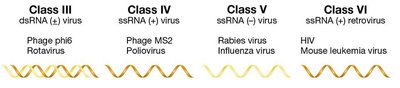
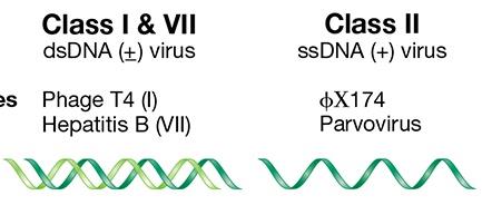
Single-stranded RNA viruses may be plus-sense (+ssRNA) (same sequence as mRNA) or minus-sense (-ssRNA) (complementary to mRNA).
Genome type determines replication strategy and is used for virus classification.
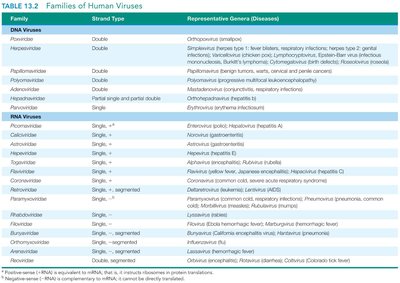
Families of Human Viruses
Human viruses are classified based on their genome type and structure. The following table summarizes major families:
Family | Strand Type | Representative Genera (Diseases) |
|---|---|---|
Parvoviridae | Single, + | Parvovirus (parvovirus) |
Herpesviridae | Double | Herpes simplex, Varicella-zoster (herpes, chickenpox) |
Picornaviridae | Single, + | Enterovirus, Rhinovirus (polio, common cold) |
Orthomyxoviridae | Single, -, segmented | Influenza virus (influenza) |
Retroviridae | Single, + | HIV (AIDS) |
Capsid Shape and Structure
Helical, Polyhedral, and Complex Capsids
Capsids are constructed from capsomere proteins and serve to protect the viral genome. The shape of the capsid is a key feature in virus classification.
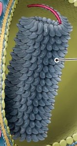
Helical: Rod-shaped, with genome coiled inside; e.g., tobacco mosaic virus.
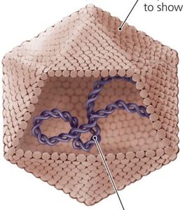
Polyhedral: Most commonly icosahedral, nearly spherical; e.g., adenovirus.
Complex: Irregular shapes, often seen in bacteriophages with additional structures for host infection.
Viral Envelopes
Structure and Function
Enveloped viruses possess a lipid bilayer surrounding their nucleocapsid, which provides protection and aids in host recognition. Proteins embedded in the envelope facilitate attachment to host cells.
Envelope: Derived from host cell membrane; contains viral glycoproteins and host proteins.
Matrix proteins: Located between envelope and nucleocapsid; stabilize virion.
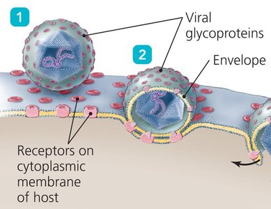
Attachment: Glycoproteins in the envelope bind to host cell receptors.
Naked viruses: Lack an envelope; consist only of nucleocapsid.
Viral Hosts and Host Specificity
Range and Mechanisms
Viruses can infect all types of living cells, including eukaryotes, bacteria, and archaea. Host specificity is determined by the ability of the virion to bind to host cell receptors.
Bacteriophages (phages): Viruses that infect bacteria.
Host specificity: Determined by viral proteins binding to specific host cell receptors.
Generalist viruses: Infect multiple hosts; others are highly specific.
Unique Features of Viruses vs Cells
Comparative Properties
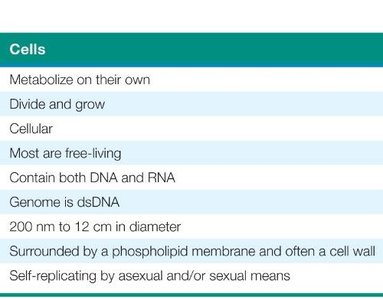
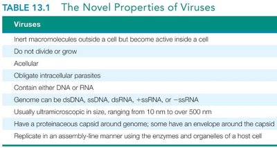
Viruses differ fundamentally from cells in their structure, replication, and metabolic capabilities. The following table summarizes these differences:
Property | Cells | Viruses |
|---|---|---|
Metabolism | Self-metabolizing | Inert outside cell |
Growth | Divide and grow | Do not grow |
Structure | Cellular | Acellular |
Genome | dsDNA | DNA or RNA (ds or ss) |
Replication | Self-replicating | Obligate intracellular |
Size | 200 nm – 12 cm | 10 nm – 500 nm |
Membrane | Phospholipid membrane | Protein capsid; sometimes envelope |
General Virus Life Cycle
Steps of Viral Replication
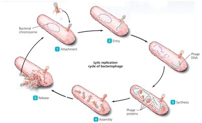
The viral life cycle consists of five main steps, each critical for the production of new virions:
Attachment: Virion binds to host cell via specific proteins.
Entry: Virion or genome enters host cell; capsid is removed if entire virion enters.
Synthesis: Host cell machinery produces viral proteins and genome copies.
Assembly: New virions are assembled from synthesized components.
Release: Virions exit host cell via lysis (bursting) or budding (enveloped viruses).
Attachment
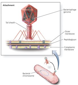
Attachment is mediated by viral proteins binding to host cell receptors. This step determines host specificity.
Capsid or envelope proteins interact with host cell surface molecules.
Example: SARS-CoV-2 spike protein binds to ACE2 receptor on lung cells.
Bacteriophages use tail fibers for attachment.
Entry
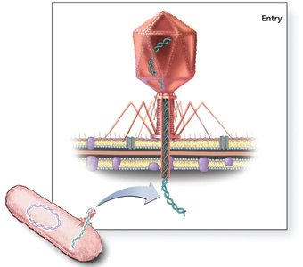
Entry involves the introduction of the viral genome into the host cell. In bacteriophages, only the genome enters, while the capsid remains outside.
Capsid proteins are removed if the entire virion enters.
Bacteriophages use lysozyme to break down bacterial cell wall and inject genome.
Host cell chromosome may be destroyed during entry.
Synthesis and Assembly
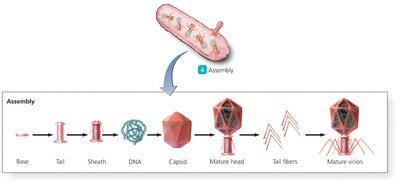
Host cell enzymes and ribosomes synthesize viral proteins and replicate the viral genome. Assembly is tightly regulated and occurs in a specific order.
Early proteins control replication; late proteins facilitate virion release.
Virion components are assembled into mature particles.
Release
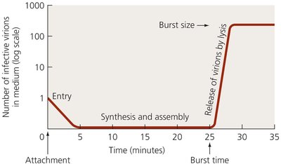
Virions are released from the host cell to infect new cells. Release can occur via lysis (cell bursts) or budding (virion acquires envelope).
Bacteriophages typically release via lysis.
Enveloped viruses release by budding, acquiring a lipid envelope.
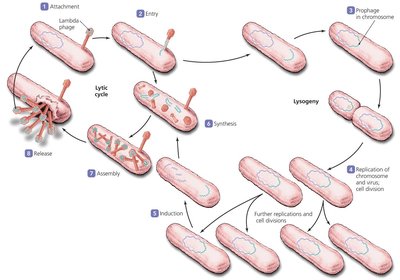
Lytic and Lysogenic Life Cycles of Bacteriophages
Lytic Cycle
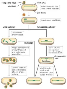
The lytic cycle involves active replication of the phage, production of new virions, and destruction of the host cell.
Phage attaches, injects genome, replicates, assembles, and lyses host cell.
All virions are released simultaneously.
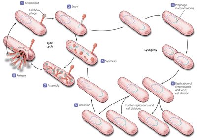
Lysogenic Cycle
Temperate phages can integrate their genome into the host cell's chromosome, becoming a prophage. The phage genome is replicated along with the host cell and can be induced to enter the lytic cycle.
Phage genome integrates into host genome.
Prophage is replicated as host divides.
Environmental signals can trigger induction into the lytic cycle.
Applications: Bacteriophages in Therapy
Phage Therapy
Bacteriophages can be used to treat bacterial infections, especially those resistant to antibiotics. Phage therapy exploits the specificity of phages for bacterial hosts.
Phages target and lyse specific bacteria.
Potential for use against antibiotic-resistant strains.
Summary Table: Acellular Infectious Agents
Agent | Structure | Genome | Replication |
|---|---|---|---|
Virus | Capsid (protein), sometimes envelope | DNA or RNA | Obligate intracellular |
Prion | Protein only | None | Induces misfolding of host proteins |
Viroid | RNA only | RNA | Obligate intracellular (plants) |
Additional info: For further study, refer to textbook sections on viral structure, genome classification, and phage life cycles. Homework and lab preparation instructions are provided in the course announcements.
