 Indietro
Acellular Infectious Agents: Viruses, Viroids, and Prions
Guida di studio - Note intelligenti
 Appunti personalizzati basati sui tuoi materiali, ampliati con definizioni chiave, esempi e contesto.
Viruses: Structure, Life Cycles, and Applications
General Virus Life Cycle
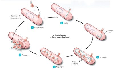
The viral replication cycle consists of five distinct steps, which are fundamental to understanding how viruses propagate within host cells. These steps are common to both bacteriophages and animal viruses, though the details may vary depending on the host and virus type.
Attachment (Adsorption): The virion binds to specific receptors on the host cell surface.
Entry: The virion or its nucleic acid enters the host cell.
Synthesis: The host cell machinery is redirected to synthesize viral nucleic acids and proteins.
Assembly: Newly synthesized viral components are assembled into complete virions.
Release: Mature virions exit the host cell, either by lysis or budding.
Lytic and Lysogenic Life Cycles of Bacteriophages
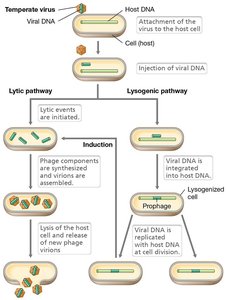
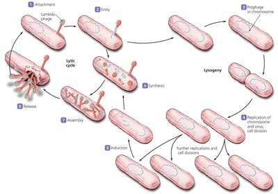
Bacteriophages can follow two main life cycles: lytic and lysogenic. The lytic cycle results in the destruction of the host cell, while the lysogenic cycle allows the phage genome to persist within the host without immediate lysis.
Lytic Cycle: The phage invades the host, replicates, and lyses the cell to release new virions.
Lysogenic Cycle: The phage genome integrates into the host genome (forming a prophage) and is replicated as the host divides. Environmental signals can induce the prophage to enter the lytic cycle.
Bacteriophages as Treatments for Bacterial Infections
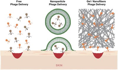
Lytic bacteriophages are being explored as alternatives to antibiotics for treating bacterial infections. Their specificity and ability to lyse bacterial cells make them promising candidates for targeted therapy.
Specificity: Phages can be selected or engineered to target specific pathogens.
Delivery Methods: Phages can be applied topically, orally, or via inhalation.
Advantages: Lytic phages rapidly destroy bacterial cells, reducing infection.
Animal Viruses: Infection Types and Life Cycle
Types of Viral Infections in Animal Cells
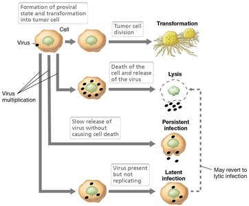
Animal viruses can cause four distinct types of infections, each with unique outcomes for the host cell.
Virulent/Lytic: Host cell is lysed to release virions.
Persistent: Host cell slowly releases virions without lysis.
Latent: Virus genome integrates into host genome and remains dormant.
Transformation: Virus induces cancerous changes in host cells.
Entry and Uncoating of Animal Viruses
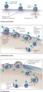
Animal viruses utilize several mechanisms to enter host cells, and uncoating is essential for viral replication.
Direct Penetration: Capsid binds to membrane, genome enters cell.
Membrane Fusion: Envelope fuses with cell membrane, nucleocapsid enters.
Endocytosis: Entire virion is internalized, nucleocapsid released from vesicle.
Uncoating: Viral genome must be released from capsid for replication.
Viral Enzymes in Animal Viruses
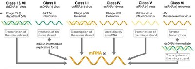
Some animal viruses require unique enzymes for replication, which are not found in host cells. These enzymes are often packaged within the virion.
RNA-dependent DNA polymerase (Reverse Transcriptase): Converts RNA to DNA in retroviruses.
RNA-dependent RNA polymerase: Synthesizes RNA from an RNA template.
Host Enzymes: Host DNA and RNA polymerases are used for DNA viruses.

Viral Budding and Virion Formation
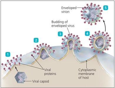
Enveloped animal viruses acquire their envelope through budding, a process that does not necessarily kill the host cell.
Budding: Nucleocapsid is wrapped in lipid membrane as it exits the cell.
Sources of Envelope: Cell membrane, endoplasmic reticulum, or nuclear membrane.
Protein-Mediated Cleavage: Some viruses require enzymes (e.g., neuraminidase in influenza) for release.
Acellular Infectious Agents: Viruses, Viroids, and Prions
Viroids: Structure and Pathogenesis
Viroids are infectious RNA molecules that lack a protein coat and do not encode proteins. They are unique to plants and cause various diseases by interfering with gene expression.
Structure: Small, circular, single-stranded RNA (239-399 nucleotides).
Replication: Use host cell machinery; cannot replicate independently.
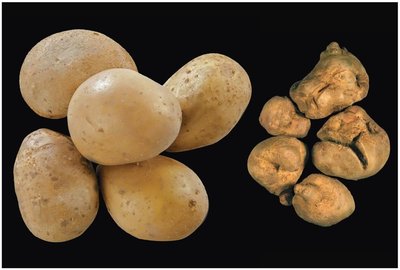
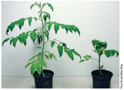
Pathogenesis: Cause growth defects, leaf and fruit malformation, and death in plants.

Prions: Infectious Proteins and Disease Mechanisms
Prions are infectious proteins that cause neurodegenerative diseases by inducing abnormal folding of normal cellular proteins. They are highly resistant to destruction and can persist in the environment.
Structure: No nucleic acid; composed solely of protein (PrP).
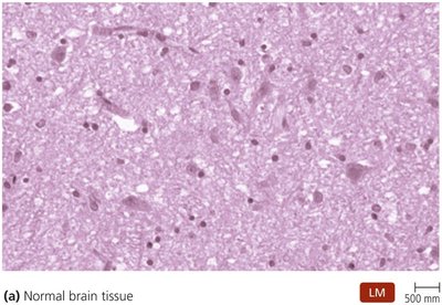
Pathogenesis: Aggregates of prion proteins kill cells, leading to spongiform encephalopathies.
Transmission: Spread depends on amino acid sequence compatibility between species.
Resistance: Prions are resistant to proteases, disinfectants, and extreme conditions.
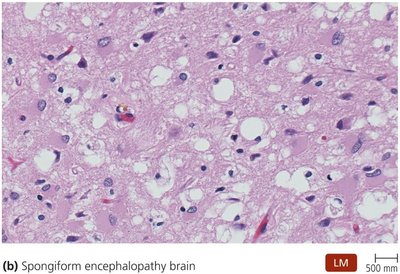
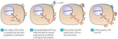
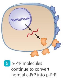

Chronic Wasting Disease (CWD) in Cervids
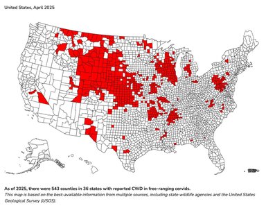
Chronic wasting disease is a prion disease affecting cervids (deer, elk, moose, etc.), with increasing prevalence across North America.
Distribution: CWD is found in saliva, urine, muscle, lymph nodes, brain, placenta, and gut tissues.
Spread: The disease is expanding geographically, as shown in recent maps.
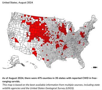
Comparison of Bacteria, Viruses, Viroids, and Prions
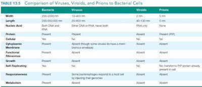
The following table summarizes the key differences between bacteria, viruses, viroids, and prions, focusing on structure, genome, and biological properties.
Property | Bacteria | Viruses | Viroids | Prions |
|---|---|---|---|---|
Width | 200–1000 nm | 10–400 nm | 2–10 nm | 5 nm |
Nucleic Acid | DNA and RNA | Either DNA or RNA | RNA | None |
Protein | Present | Present | Absent | Present (PrP) |
Cytoplasmic Membrane | Present | Absent (some viruses have envelope) | Absent | Absent |
Functional Ribosomes | Present | Absent | Absent | Absent |
Growth | Present | Absent | Absent | Absent |
Self-Replicating | Present | Some bacteriophages respond to host cell | Absent | No; transforms PrP protein already present in cell |
Metabolism | Present | Absent | Absent | Absent |
Laboratory Techniques: Plaque Assays
Plaque Assays for Virus Quantification
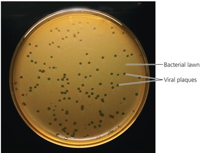
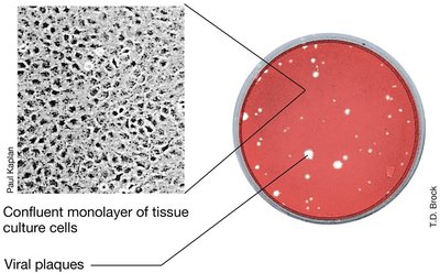
Plaque assays are used to quantify viral titers in both bacteriophages and animal viruses. The formation of plaques indicates sites of infection and allows calculation of viral concentration.
Plaque-Forming Units (PFU): Viral particles are quantified as PFU/mL.
Applications: Used for research, diagnostics, and vaccine production.
Summary Table: Acellular Infectious Agents
Key Differences and Properties
This table provides a concise comparison of the main acellular infectious agents and bacteria, highlighting their structural and functional differences.
Agent | Genome | Protein Coat | Host Range | Replication |
|---|---|---|---|---|
Bacteria | DNA & RNA | Yes | Many | Self-replicating |
Virus | DNA or RNA | Yes | Specific | Host-dependent |
Viroid | RNA | No | Plants | Host-dependent |
Prion | None | No | Animals | Protein conversion |
Additional info: Expanded explanations and context were added to ensure completeness and clarity for exam preparation.
