 Indietro
Aseptic Technique and Colony Morphology in Microbiology Lab
Guida di studio - Note intelligenti
 Appunti personalizzati basati sui tuoi materiali, ampliati con definizioni chiave, esempi e contesto.
Microbes in the Environment: Aseptic Technique and Colony Morphology
Introduction
This guide covers essential microbiology laboratory skills, focusing on aseptic technique for transferring bacteria and the observation and classification of microbial colonies. Mastery of these techniques is foundational for preventing contamination and accurately studying microorganisms.
Colony Morphology: Reading Your Plates
Whole-Colony Appearance
Microbial colonies can be distinguished by their visible characteristics on solid media. Careful observation and recording of these features are crucial for identification and comparison.
Shape (Form): The overall outline of the colony when viewed from above.
Margin: The edge or border of the colony.
Elevation: The side profile or height of the colony above the agar surface.
Pigment (Color): The color produced by the colony, which may be due to pigments secreted by the microorganism.
Size: Measured as the diameter in millimeters (mm).
Number: The exact count of colonies of each type. If too numerous to count, record as "TNTC" (Too Numerous To Count).
Important Note: If a plate shows fuzzy, cottony, or web-like growth (indicative of fungi), do not open the lid. Observe and describe from outside the plate.
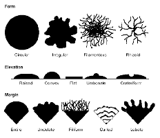
Describing Colony Morphology
Form (Shape):
Circular: Round and uniform.
Irregular: Non-uniform, uneven edges.
Filamentous: Thread-like extensions.
Rhizoid: Root-like, branching appearance.
Margin (Edge):
Entire: Smooth, even edge.
Undulate: Wavy edge.
Lobate: Lobed, irregular projections.
Filamentous: Thread-like extensions from the edge.
Elevation (Profile):
Flat: No noticeable elevation.
Raised: Slightly elevated above the agar.
Convex: Dome-shaped, rounded.
Umbonate: Raised with a central bump.
Example: A colony described as "circular, entire, convex, yellow pigment, 2 mm diameter, 15 colonies" provides a comprehensive morphological profile.
Aseptic Technique: Transfer of Bacteria
Purpose of Aseptic Technique
Aseptic technique is a set of practices used to prevent contamination of cultures, media, and the laboratory environment. It is essential for:
Protecting the culture: Prevents introduction of unwanted microbes into pure cultures.
Protecting the experimenter: Reduces the risk of exposure to potentially harmful organisms.
General Protocol for Aseptic Transfer
Label all tubes: Include your name, date, organism name, and type of medium (broth, slant, or deep).
Sterilize the loop or needle: Hold in the flame until red-hot; allow to cool without touching any surface.
Open tubes aseptically: Uncap both stock and blank tubes, flaming the mouth of each before and after opening.
Transfer using the correct technique: Use the appropriate tool and method for each medium (see below).
Flame, recap, and re-sterilize: Flame tube mouths again, recap, and sterilize the loop/needle before setting it down.
Repeat for each medium: Use the same careful technique for all transfers.
Specific Transfer Techniques
Medium | Tool | Transfer Method |
|---|---|---|
Broth | Loop | Dip sterile loop into stock broth, then swirl into new broth tube to mix sample. |
Slant | Loop | Starting at the bottom, drag loop in a tight zigzag up the agar surface. |
Deep | Needle | Insert straight needle down the center of the agar, almost to the bottom, then withdraw along the same path. |
After the Lab: Clean-Up and Record-Keeping
Record all observations and results for each colony type (diameter, appearance, margin, elevation, pigment, count).
Document all inoculations performed (broth, slant, deep) and label tubes for incubation.
Clean your workspace: Wipe down surfaces and place inoculation tubes in racks.
Looking Ahead
Read about smear preparation and simple staining for the next class.
Complete any assigned quizzes and ensure your lab notebook is up to date.
Additional info: Mastery of aseptic technique and colony morphology is foundational for all subsequent microbiology lab work, including identification, isolation, and characterization of microorganisms.
