 Indietro
Chapter 4: Bacteria and Archaea: Structure, Function, and Classification- Book Notes
Guida di studio - Note intelligenti
 Appunti personalizzati basati sui tuoi materiali, ampliati con definizioni chiave, esempi e contesto.
Chapter 4: Bacteria and Archaea
Section 4.1: The Bacteria
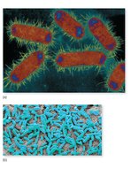
Bacteria are single-celled prokaryotic organisms capable of performing all essential life processes. They exhibit a wide variety of shapes, sizes, and arrangements, and can exist as individual cells or in complex communities such as biofilms.
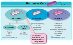
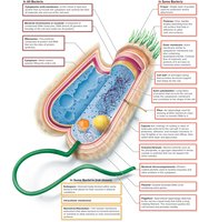
Structures all bacteria possess: Cytoplasmic membrane, cytoplasm, ribosomes, and a chromosome (nucleoid).
Structures found in some bacteria: Cell wall, glycocalyx (capsule or slime layer), flagella, pili, fimbriae, endospores, plasmids, and inclusion bodies.
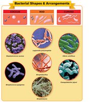
Major shapes of bacteria:
Coccus: Spherical, oval, or bean-shaped.
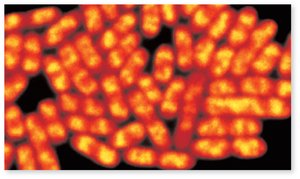
Bacillus: Rod-shaped, can be blocky, spindle-shaped, or filamentous.
Curved: Includes vibrio (comma-shaped), spirillum (rigid spiral), and spirochete (flexible corkscrew).
Pleomorphism: Variation in size and shape within a single species due to genetic or environmental factors.
Bacterial arrangements: Diplococci (pairs), tetrads (groups of four), staphylococci (clusters), streptococci (chains), sarcina (cubical packets), diplobacilli (pairs of rods), streptobacilli (chains of rods), palisades (side-by-side rods).
Biofilms
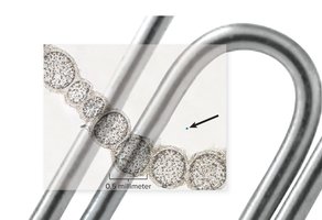
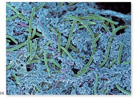
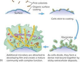
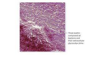
Biofilms are structured communities of bacteria and other microorganisms attached to surfaces and embedded in a self-produced extracellular matrix. They provide protection and enhanced survival for their inhabitants.
Section 4.2: External Structures
Bacterial external structures include appendages for motility and attachment, as well as surface layers for protection and interaction with the environment.
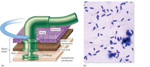
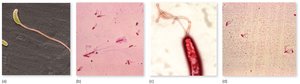
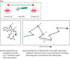
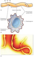
Flagella: Long, whip-like structures used for motility. Arrangements include monotrichous (single), lophotrichous (tufts), amphitrichous (both ends), and peritrichous (all over surface).
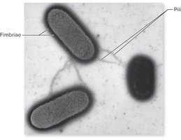
Fimbriae: Short, bristle-like fibers for attachment to surfaces and other cells.
Pili: Longer, tubular structures involved in DNA transfer (conjugation) and attachment.
Nanowires: Thin, tubular extensions for electron and nutrient transfer between cells.
S layer: Protective protein layer produced in hostile environments.
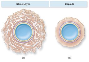
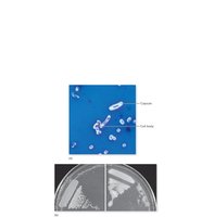
Glycocalyx: Polysaccharide coating that can be a loose slime layer or a dense capsule, providing protection and aiding in biofilm formation.
Section 4.3: The Cell Envelope
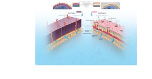
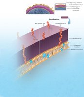
The bacterial cell envelope is a multi-layered structure that protects the cell and mediates interactions with the environment. It consists of the cell wall, cytoplasmic membrane, and, in some bacteria, an outer membrane.
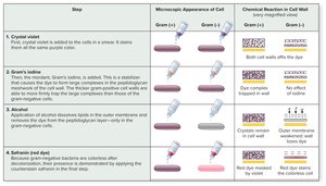
Gram-positive bacteria: Thick peptidoglycan cell wall, teichoic acids, and a single cytoplasmic membrane.
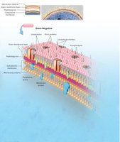
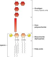
Gram-negative bacteria: Thin peptidoglycan layer, outer membrane containing lipopolysaccharide (LPS), and a cytoplasmic membrane.
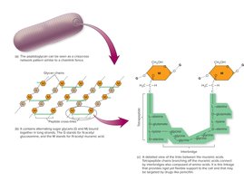
Peptidoglycan: A mesh-like polymer of sugars and amino acids providing structural support.
Lipopolysaccharide (LPS): Found in the outer membrane of gram-negative bacteria; acts as an endotoxin.
Porins: Proteins in the outer membrane that regulate molecule entry.
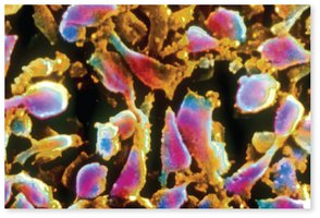
Nontypical cell walls: Mycobacterium and Nocardia have waxy mycolic acids; Mycoplasmas lack cell walls entirely.
Section 4.4: Bacterial Internal Structure
Bacterial cytoplasm contains the genetic material, ribosomes, storage granules, and other structures necessary for cellular function and survival.
Nucleoid: Region containing the single, circular bacterial chromosome.
Plasmids: Small, nonessential DNA molecules that confer advantages such as antibiotic resistance.
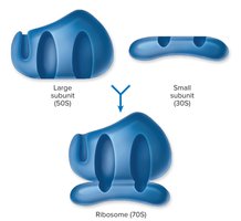
Ribosomes: Sites of protein synthesis; bacterial ribosomes are 70S (composed of 50S and 30S subunits).
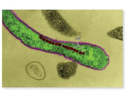
Inclusion bodies: Storage sites for nutrients and other substances.
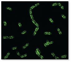
Cytoskeleton: Protein filaments that help maintain cell shape.
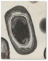
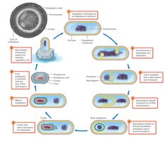
Endospores: Highly resistant, dormant structures formed by some bacteria (e.g., Bacillus, Clostridium) in response to adverse conditions.
Section 4.5: The Archaea
Archaea are prokaryotes distinct from bacteria, with unique genetic, biochemical, and structural features. They often inhabit extreme environments.
Differences from bacteria: Unique rRNA sequences, membrane lipids, cell wall composition (lack true peptidoglycan), and DNA compaction methods.
Types of archaea:
Methanogens: Produce methane from CO2 and H2.
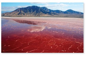
Extreme halophiles: Require high salt concentrations.
Psychrophiles: Grow at low temperatures.
Hyperthermophiles: Grow at very high temperatures.
Section 4.6: Classification Systems for Bacteria and Archaea
Bacteria and archaea are classified based on phenotypic and genetic characteristics. Bergey’s Manuals are the primary references for identification and classification.
Bergey’s Manual of Systematic Bacteriology: Comprehensive, uses phenotypic and genetic data.
Bergey’s Manual of Determinative Bacteriology: Focuses on phenotypic traits for identification in clinical and teaching labs.
Major divisions:
Gracilicutes: Gram-negative cell walls.
Firmicutes: Gram-positive cell walls.
Tenericutes: Lack cell walls.
Mendosicutes: Archaea.
Species and subspecies: Bacterial species are groups of strains with similar traits; subspecies (strains, types, serotypes) differ in minor characteristics or antigenic properties.

Additional info: This guide covers the essential structural, functional, and classification features of bacteria and archaea, integrating visual aids for enhanced understanding. For further study, refer to Bergey’s Manuals and laboratory identification protocols.
