 Indietro
Bacterial Motility and Flagella: Laboratory Techniques and Structural Arrangements
Guida di studio - Note intelligenti
 Appunti personalizzati basati sui tuoi materiali, ampliati con definizioni chiave, esempi e contesto.
Bacterial Motility
Introduction to Bacterial Motility
Bacterial motility refers to the ability of bacteria to move independently, often using specialized structures such as flagella. Motility is a key characteristic for identification and understanding of bacterial behavior, especially in clinical and environmental microbiology.
Motility is defined as self-propelled movement over relatively long distances, distinguishing it from random vibrations (Brownian motion) or passive movement caused by water currents.
Motile bacteria can navigate toward nutrients or away from harmful substances (chemotaxis).
Common motile bacteria include Escherichia coli and Pseudomonas species.
Non-motile bacteria, such as Staphylococcus epidermidis, lack these movement capabilities.
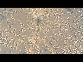
Hanging Drop Slide Technique
Principle and Purpose
The hanging drop slide is a classic microbiological technique used to observe live bacteria and assess their motility. This method allows for the visualization of true motility, as opposed to Brownian motion or movement caused by currents in the medium.
True motility is characterized by directional movement of cells across the field of view.
Brownian motion refers to random, non-directional vibrations caused by collisions with water molecules.
Water currents may move cells passively but do not indicate motility.
Hanging Drop Slide Preparation Procedure
Proper preparation of the hanging drop slide is essential for accurate observation of bacterial motility.
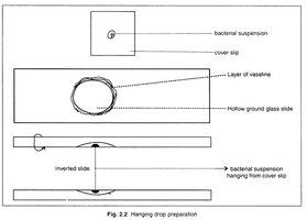
Apply a thin ring of petroleum jelly (Vaseline) around the well of a depression (hollow ground) slide.
Place a small drop of bacterial suspension (e.g., E. coli) on a clean coverslip.
Invert the prepared depression slide over the coverslip, centering the drop under the well.
Press gently to seal the coverslip to the slide, creating an airtight chamber.
Observe the preparation under oil immersion (1000X) using a microscope. Reduce light with the iris diaphragm for better contrast.
Repeat the procedure with a non-motile control, such as S. epidermidis.
Post-Observation Cleanup
Separate the coverslip from the slide and soak in a staining tray with bleach for 15 minutes to disinfect.
Dispose of the coverslip in glass waste.
Rinse the depression slide with ethanol (Gram’s decolorizer) and water, then return to the slide tray for reuse.
Flagella
Structure and Visualization
Flagella are long, thin, whip-like appendages that provide motility to many bacterial species. They are typically too thin to be seen with simple staining techniques and require special methods for visualization.
Flagella are composed of the protein flagellin and are anchored in the cell membrane and wall.
Special stains (e.g., flagella stain) or electron microscopy are used to visualize flagella.
Flagellar Arrangements
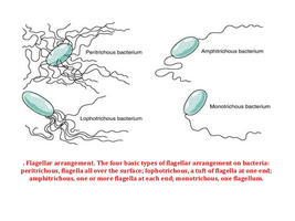
Bacteria can be classified based on the number and arrangement of their flagella, which is important for identification and understanding motility patterns.
Arrangement | Description | Example |
|---|---|---|
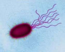
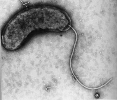
Monotrichous | Single flagellum at one end | Vibrio cholerae |
Amphitrichous | Single flagellum at both ends | Alcaligenes faecalis |
Lophotrichous | Tuft of flagella at one end | Pseudomonas fluorescens |
Peritrichous | Flagella distributed all over the cell surface | Escherichia coli |
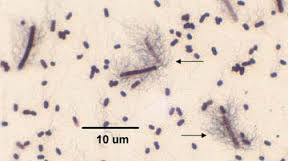
Microscopic Observation of Flagella
In laboratory settings, prepared slides stained specifically for flagella are examined under high magnification (1000X) to identify the arrangement and presence of flagella.
Flagella stains increase the diameter of flagella, making them visible under the light microscope.
Identification of flagellar arrangement aids in bacterial classification and diagnosis.
Key Points for Laboratory Practice
Always reduce the light reaching the sample with the iris diaphragm and/or light control to enhance contrast when observing live, unstained bacteria.
Proper disposal and cleaning of slides and coverslips are essential for laboratory safety and reuse of materials.
Understanding motility and flagellar arrangements is fundamental for bacterial identification and taxonomy.
