 Indietro
Biochemical and Hemolytic Testing of Gram-Positive Bacteria: Microbiology Lab Study Guide
Guida di studio - Note intelligenti
 Appunti personalizzati basati sui tuoi materiali, ampliati con definizioni chiave, esempi e contesto.
Biochemical Testing of Gram-Positive Bacteria
Hydrolysis: Definitions and Mechanisms
Hydrolysis is a fundamental biochemical reaction in microbiology, involving the splitting of large molecules into smaller units through the addition of water. This process is catalyzed by hydrolytic enzymes, which can act either inside the cell (intracellular) or outside the cell (extracellular).
Intracellular hydrolysis: The reaction occurs within the cell. Examples include the bile esculin and urea tests.
Extracellular hydrolysis: Enzymes are secreted and act outside the cell. Examples include starch, gelatin, and DNA hydrolysis tests.
Hydrolytic enzymes: These enzymes facilitate the breakdown of complex molecules, aiding in nutrient acquisition and identification of microbial species.
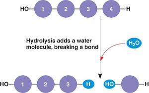
Additional info: Hydrolysis is essential for microbial metabolism and is used diagnostically to differentiate bacterial species based on their enzymatic capabilities.
DNA Hydrolysis Test
The DNA hydrolysis (DNase) test is a differential medium used to distinguish among certain genera of both Gram-positive and Gram-negative bacteria. The medium contains peptides for growth, NaCl for osmotic balance, DNA as the substrate, and methyl green as an indicator.
Principle: Methyl green binds to intact DNA, giving the medium a green color. When DNA is hydrolyzed, methyl green is released, resulting in a colorless zone around colonies that produce DNase.
Interpretation: A clear zone around the bacterial growth indicates DNase activity (positive test).
Application: Used to differentiate Staphylococcus aureus (DNase positive) from other staphylococci and related genera.
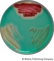
Additional info: The DNase test is important for identifying pathogenic bacteria that can degrade host DNA as a virulence factor.
Antimicrobial Susceptibility Testing
Principles of Disk Diffusion Method
Antimicrobial susceptibility testing determines the sensitivity or resistance of bacteria to specific antibiotics. The disk-diffusion method (Kirby-Bauer test) is commonly used in clinical microbiology.
Procedure: Antibiotic-impregnated disks are placed on an agar plate inoculated with the test organism. After incubation, zones of inhibition (areas with no bacterial growth) are measured.
Interpretation: The diameter of the zone is compared to standardized charts to determine susceptibility or resistance. The presence of a zone alone does not indicate susceptibility; the size is critical.
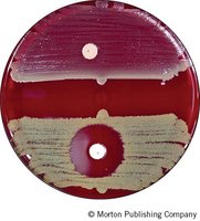
Additional info: This method is essential for guiding clinical therapy and for epidemiological surveillance of resistance.
Bacitracin Susceptibility Test
Bacitracin is an antibiotic that inhibits cell wall synthesis. This test is used to differentiate β-hemolytic group A streptococci (GAS), especially Streptococcus pyogenes (sensitive), from other β-hemolytic streptococci and to distinguish Staphylococcus (resistant) from Micrococcus (sensitive).
Interpretation: A zone of inhibition ≥10 mm indicates susceptibility.
Application: Rapid identification of S. pyogenes in clinical specimens.
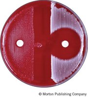
Novobiocin Susceptibility Test
Novobiocin interferes with bacterial ATPase activity. This test is primarily used to differentiate coagulase-negative staphylococci, especially to identify Staphylococcus saprophyticus (novobiocin resistant, a common cause of urinary tract infections).
Interpretation: A zone of inhibition ≥16 mm indicates susceptibility.
Application: Differentiates S. saprophyticus (resistant) from S. epidermidis (susceptible).
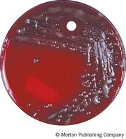
Optochin (P-disk) Susceptibility Test
Optochin also interferes with ATPase and is used to differentiate Streptococcus pneumoniae (sensitive) from other α-hemolytic streptococci (resistant).
Interpretation: A zone of inhibition ≥14 mm indicates susceptibility.
Application: Rapid identification of S. pneumoniae in respiratory specimens.
Hemolysis on Blood Agar
Types of Hemolysis
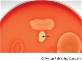
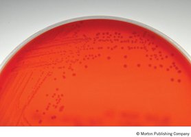
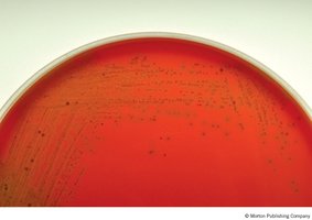
Blood agar is used to detect hemolysins produced by Gram-positive cocci. Hemolysins are enzymes that lyse red blood cells, and their activity is classified as follows:
α-hemolysis: Partial destruction of red blood cells, producing a greenish discoloration around colonies (e.g., Streptococcus pneumoniae).
β-hemolysis: Complete destruction of red blood cells, resulting in a clear zone around colonies (e.g., Streptococcus pyogenes).
γ-hemolysis: No hemolysis (nonhemolytic), with no change in the medium (e.g., Enterococcus faecalis).
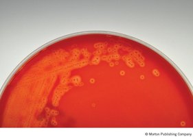
Streptolysins: These are hemolysins produced by streptococci, with types O and S being maximally effective under anaerobic conditions.
Additional info: The streak-stab technique is used to enhance detection of streptolysin activity by creating anaerobic conditions in the agar.
Coagulase Test
Principle and Applications
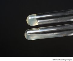
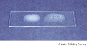
The coagulase test detects the presence of the enzyme coagulase, which enables Staphylococcus aureus to clot plasma, providing protection from host defenses. Coagulase exists in free (released) and bound (cell-associated) forms.
Media: Rabbit plasma treated with anticoagulant is used for the test.
Tube test: Detects both free and bound coagulase.
Slide test: Detects only bound coagulase (clumping factor).
Interpretation: Clot formation or visible agglutination indicates a positive result.
Application: Differentiates S. aureus (coagulase positive) from other Gram-positive cocci.
Additional info: The coagulase test is a critical diagnostic tool for identifying pathogenic staphylococci in clinical microbiology.
Laboratory Procedures Overview
Summary of Key Laboratory Activities
Inoculation of DNase plates with Staphylococcus aureus, Serratia marcescens, and Enterobacter aerogenes for DNA hydrolysis testing.
Preparation of blood agar plates for antimicrobial susceptibility testing (bacitracin, novobiocin, optochin) using appropriate bacterial cultures.
Throat culture collection and streak-stab technique for hemolysis observation.
Inoculation of coagulase tubes and performance of coagulase slide tests to differentiate S. aureus from S. epidermidis.
Additional info: These laboratory techniques are foundational for the identification and characterization of clinically significant Gram-positive bacteria.
