 Indietro
Cell Membrane & Transport: Structure, Function, and Mechanisms
Guida di studio - Note intelligenti
 Appunti personalizzati basati sui tuoi materiali, ampliati con definizioni chiave, esempi e contesto.
Cell Membrane & Transport
Cell Envelope & Biological Membranes
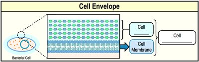
The cell envelope refers to all layers that surround the cell, including the cell membrane and, in many cases, the cell wall. The composition of the cell envelope varies among different cell types, but all cells possess a cell membrane as a fundamental component.
Cell Envelope: Includes the cell membrane and, in some organisms, additional layers such as the cell wall.
Cell Membrane: A universal structure present in all cells, forming the boundary between the cell interior and its environment.
Biological Membranes: Structure and Composition
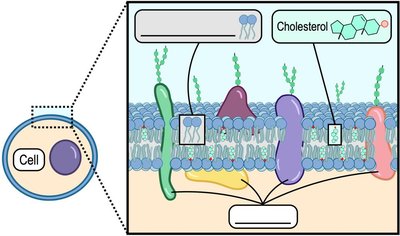
Biological membranes are primarily composed of phospholipids, which are amphipathic molecules, meaning they have both hydrophilic (water-attracting) and hydrophobic (water-repelling) regions. These membranes also contain proteins and, in some cases, cholesterol, contributing to their structure and function.
Phospholipid Bilayer: The fundamental structure of biological membranes, with hydrophilic heads facing outward and hydrophobic tails inward.
Fluid Mosaic Model: Describes membranes as dynamic, with proteins and lipids moving laterally within the bilayer.
Embedded Molecules: Proteins and cholesterol are interspersed within the bilayer, contributing to membrane fluidity and function.
Phospholipid Structure
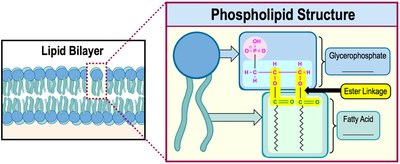
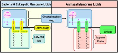
Phospholipids consist of a glycerophosphate head and two fatty acid tails, connected by ester linkages in bacteria and eukaryotes. Their amphipathic nature is crucial for forming bilayers in aqueous environments.
Hydrophilic Head: Glycerophosphate group interacts with water.
Hydrophobic Tails: Fatty acids avoid water, forming the membrane's interior.
Ester Linkage: Connects the head group to the fatty acid tails in bacteria and eukaryotes.
Bacterial vs. Eukaryotic Cell Membranes
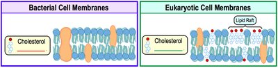
Bacterial and eukaryotic membranes differ in their lipid composition. Eukaryotic membranes contain cholesterol, which increases rigidity and forms lipid rafts, while bacterial membranes typically lack cholesterol.
Bacterial Membranes: Lack cholesterol, generally more fluid.
Eukaryotic Membranes: Contain cholesterol and lipid rafts, contributing to membrane stability and compartmentalization.
Archaeal Cell Membranes
Archaeal membranes are distinct from bacterial and eukaryotic membranes in two major ways:
Isoprene Chains: Archaeal hydrophobic tails are composed of repeating isoprene units, not fatty acids.
Ether Linkages: Archaeal lipids are connected via ether bonds, which are more resistant to heat and chemical damage than ester linkages.
Types of Archaeal Membrane Lipids
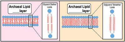
Bilayers: Formed by glycerol diether lipids, with two isoprene chains attached to a glycerophosphate head.
Monolayers: Formed by diglycerol tetraether lipids, with long isoprene chains connecting two head groups, increasing rigidity in extreme environments.
Types of Membrane Proteins
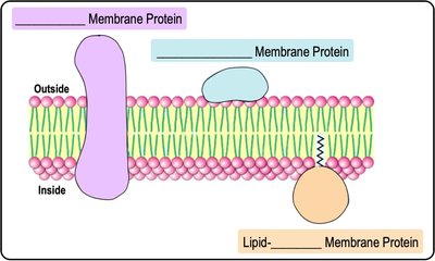
Membrane proteins are classified based on their association with the lipid bilayer:
Integral Proteins: Span the entire membrane and are embedded within the lipid bilayer.
Peripheral Proteins: Attached to the membrane surface, not embedded within the bilayer.
Lipid-Anchored Proteins: Covalently attached to lipid molecules within the membrane.
Functions of Membrane Proteins
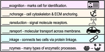
Recognition: Mark cells for identification.
Anchorage: Anchor the cytoskeleton and extracellular matrix.
Transduction: Act as receptors for signal molecules.
Transport: Facilitate molecular movement across the membrane.
Linkage: Connect cells via protein linkages.
Enzymatic Activity: Catalyze various biochemical reactions.
Membrane Transport Mechanisms
Concentration Gradients & Diffusion
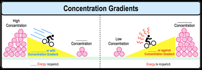
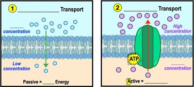
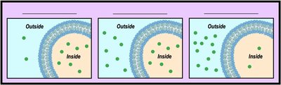
A concentration gradient is the difference in the concentration of a substance between two regions. Molecules naturally move down their concentration gradient (from high to low concentration) via diffusion, a passive process that does not require energy.
Down the Gradient: Movement from high to low concentration (no energy required).
Against the Gradient: Movement from low to high concentration (energy required).
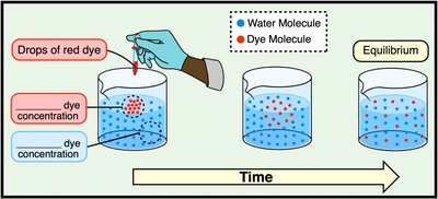
Diffusion Example
When a dye is added to water, its molecules move from an area of high concentration to low concentration until equilibrium is reached.
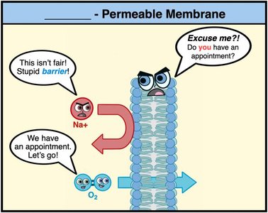
Selective Permeability of Biological Membranes
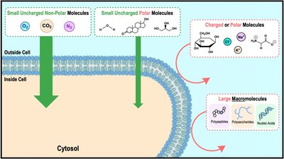
Biological membranes are selectively permeable, meaning they regulate what substances can cross. Small, nonpolar molecules can diffuse freely, while large or charged molecules require assistance.
Freely Diffusing Molecules: Small, uncharged, nonpolar (e.g., O2, CO2).
Restricted Molecules: Large, polar, or charged molecules (e.g., ions, glucose).

Overview of Membrane Transport Types
Membrane transport is divided into passive (no energy required) and active (energy required) mechanisms. Bulk transport processes such as endocytosis and exocytosis move large molecules.
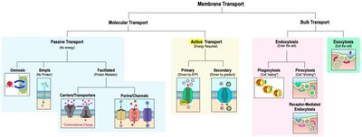
Passive vs. Active Transport
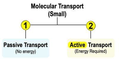
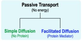
There are two main types of molecular transport across membranes:
Passive Transport: Moves substances down their concentration gradient without energy input.
Active Transport: Moves substances against their concentration gradient, requiring energy (usually ATP).
Classes of Membrane Transport Proteins
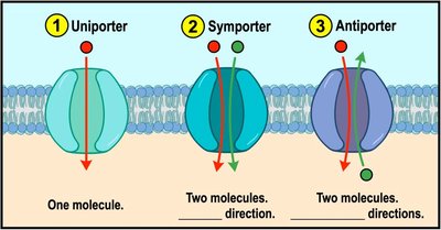
Uniporters: Transport one molecule at a time in one direction.
Symporters: Cotransport two or more molecules in the same direction.
Antiporters: Cotransport two or more molecules in opposite directions.
Osmosis and Tonicity
Osmosis
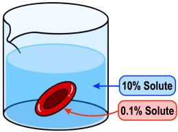
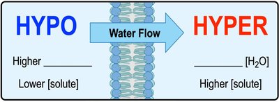
Osmosis is the passive diffusion of water across a semi-permeable membrane. The direction of water movement depends on the relative concentration of solutes (tonicity) on either side of the membrane.
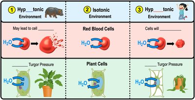
Hypotonic Solution: Lower solute concentration outside the cell; water enters the cell.
Isotonic Solution: Equal solute concentrations; water moves in and out at equal rates.
Hypertonic Solution: Higher solute concentration outside the cell; water exits the cell.
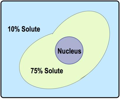
Effects of Tonicity on Cells
Hypotonic Environment: Water enters cells, causing swelling and possible lysis (bursting). Preferred by plant cells due to turgor pressure.
Isotonic Environment: Water enters and exits at equal rates; preferred by animal cells.
Hypertonic Environment: Water exits cells, causing shrinkage (crenation in animal cells, plasmolysis in plant cells).
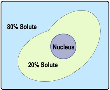
Simple & Facilitated Diffusion
Simple Diffusion
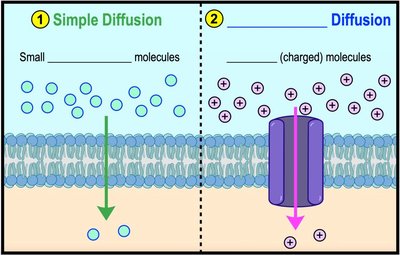
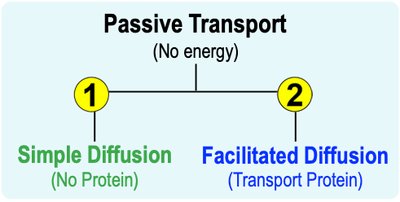
Simple diffusion is the direct movement of small, uncharged molecules across the cell membrane, down their concentration gradient, without the need for energy or transport proteins.
Facilitated Diffusion
Facilitated diffusion is the passive movement of molecules across the membrane via specific transport proteins. This process is essential for charged or larger polar molecules that cannot diffuse freely through the lipid bilayer.
Porins/Channels: Form pores for molecules to pass through (e.g., aquaporins for water).
Transporters/Carriers: Undergo conformational changes to move molecules across the membrane.
Active Transport
Primary Active Transport
Primary active transport uses energy directly from ATP hydrolysis to move molecules against their concentration gradient. This process is vital for maintaining cellular ion balances.
Example: Sodium-potassium pump (Na+/K+ ATPase) exports 3 Na+ ions and imports 2 K+ ions per ATP hydrolyzed.
Secondary Active Transport
Secondary active transport uses the energy stored in concentration gradients established by primary active transport to move other molecules against their gradients. This process does not use ATP directly but relies on the gradient created by ATP-driven pumps.
ABC Transporters
ATP-Binding Cassette (ABC) transporters are integral membrane proteins that use ATP hydrolysis to transport various substances across membranes, often against their concentration gradients. They play a key role in multidrug resistance in both bacteria and humans.
Group Translocation
Group translocation is a unique transport mechanism in prokaryotes where a molecule is chemically modified as it is transported into the cell, typically by the addition of a phosphate group. This process allows the cell to import nutrients efficiently and maintain concentration gradients.
Bulk Transport: Endocytosis & Exocytosis
Endocytosis
Endocytosis is the process by which cells engulf large molecules or particles by enclosing them in vesicles derived from the plasma membrane. There are three main types:
Phagocytosis: Engulfment of large solid particles ('cell eating').
Pinocytosis: Uptake of extracellular fluid ('cell drinking').
Receptor-Mediated Endocytosis: Specific uptake of molecules via receptor proteins.
Exocytosis
Exocytosis is the process by which cells expel large molecules or waste products by fusing vesicles with the plasma membrane, releasing their contents to the extracellular space. This is essential for secretion of hormones, neurotransmitters, and digestive enzymes.
*Additional info: This guide covers the structure and function of cell membranes, the differences among bacterial, archaeal, and eukaryotic membranes, and the mechanisms of molecular and bulk transport across membranes, as relevant to microbiology students.*
