 Indietro
Cell Structure and Function: Prokaryotic and Eukaryotic Cells
Guida di studio - Note intelligenti
 Appunti personalizzati basati sui tuoi materiali, ampliati con definizioni chiave, esempi e contesto.
Cell Structure and Function
Introduction
This chapter explores the fundamental differences and similarities between prokaryotic and eukaryotic cells, focusing on their structures, functions, and the molecular mechanisms that underlie cellular processes. Understanding these basics is essential for all microbiology students, as cell structure is foundational to microbial physiology, genetics, and pathogenesis.
Features of Prokaryotic Cells
General Characteristics
Prokaryotes include Bacteria and Archaea.
They lack a membrane-bound nucleus and most internal membrane-bound organelles.
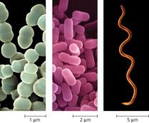
Typically small (~1.0 μm in diameter) and structurally simple.
Genetic material is located in a region called the nucleoid.
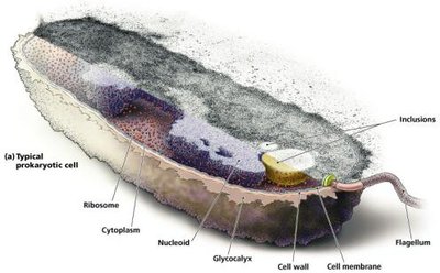
External Structures of Prokaryotes
Glycocalyces: Gelatinous, sticky substances surrounding the cell, composed of polysaccharides, polypeptides, or both. They protect against desiccation and immune recognition.
Two types:
Capsule: Firmly attached to the cell surface, often enhancing pathogenicity by evading the immune system.
Slime Layer: Loosely attached, aids in surface attachment and biofilm formation.
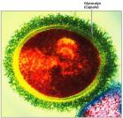
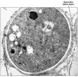
Flagella
Flagella are long, whip-like appendages responsible for motility in many prokaryotes. They are complex molecular machines that convert rotary motion into directional movement.
Composed of three main parts: filament (flagellin protein), hook, and basal body.
The basal body anchors the flagellum to the cell wall and membrane.
Flagella rotate 360°, enabling movement toward or away from stimuli (taxis).
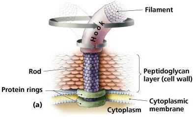
Arrangements of Flagella
Monotrichous: Single flagellum at one end.
Lophotrichous: Cluster of flagella at one end.
Amphitrichous: Flagella at both ends.
Peritrichous: Flagella distributed over the entire cell surface.
Endoflagella: Found in spirochetes, enabling corkscrew motion.
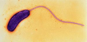
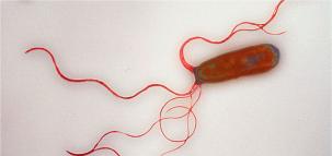
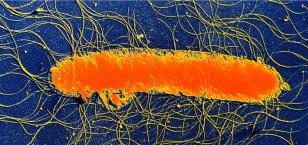
Flagellar Function
Movement is achieved by rotation, which can be clockwise or counterclockwise.
Runs: Straight movement, increased by positive stimuli (chemotaxis/phototaxis).
Tumbles: Random changes in direction, increased by negative stimuli.
Fimbriae and Pili
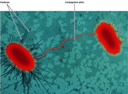
Fimbriae: Short, bristle-like projections used for attachment to surfaces, other cells, and in biofilm formation.
Pili (conjugation pili/sex pili): Longer than fimbriae, usually 1-2 per cell, mediate DNA transfer between cells (conjugation).
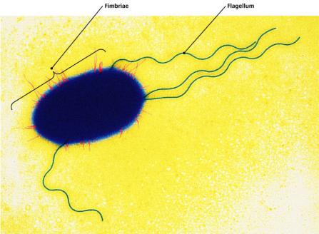
Prokaryotic Cell Walls
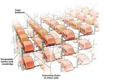
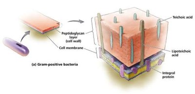
The cell wall provides structural support, shape, and protection from osmotic pressure. Most bacterial cell walls are composed of peptidoglycan.
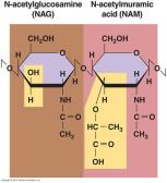
Peptidoglycan: A lattice of polysaccharide chains (N-acetylglucosamine [NAG] and N-acetylmuramic acid [NAM]) cross-linked by short peptides.
Gram-Positive vs. Gram-Negative Cell Walls
Feature | Gram-Positive | Gram-Negative |
|---|---|---|
Peptidoglycan Layer | Thick (~40 layers) | Thin (~10 layers) |
Outer Membrane | Absent | Present (contains LPS) |
Gram Stain | Purple (crystal violet) | Pink (safranin) |
Teichoic Acids | Present | Absent |
Prokaryotic Cell Membrane
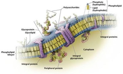
Composed of a phospholipid bilayer with embedded proteins (integral, peripheral, glycoproteins).
Described by the fluid mosaic model.
Functions: Selective permeability, energy production, and transport.
Transport Across the Membrane
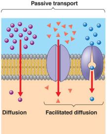
Passive Transport: No energy required; includes simple diffusion, osmosis, and facilitated diffusion.
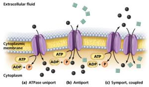
Active Transport: Requires ATP; moves substances against their concentration gradient using uniporters, antiporters, and symporters.
Cytoplasm of Prokaryotes
Includes cytosol (liquid), inclusions (storage granules), ribosomes (protein synthesis), cytoskeleton (cell shape), enzymes, and genetic material (DNA/RNA).
Features of Eukaryotic Cells
General Characteristics
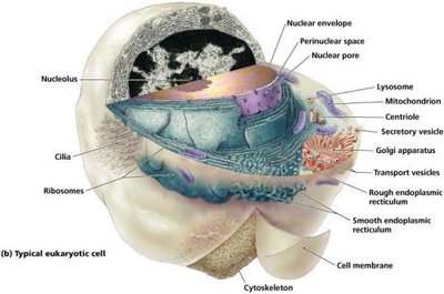
Larger (10–100 μm), more complex, with a true nucleus and internal membrane-bound organelles.
Includes algae, protozoa, fungi, animals, and plants.
External Structures of Eukaryotes
Extracellular Matrix (ECM): Anchors cells, strengthens surfaces, protects against dehydration, and mediates cell recognition and communication.
Flagella and Cilia: Used for movement; structurally distinct from prokaryotic flagella.
Flagella and Cilia Structure
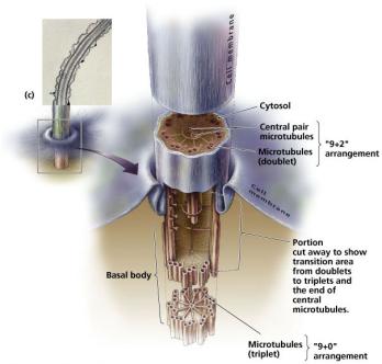
Eukaryotic flagella have a "9 + 2" arrangement of microtubules (tubulin protein) and lack a hook.
Basal body has a "9 + 0" arrangement.
Movement is undulatory (wave-like), not rotational.
Flagella: Prokaryotes vs. Eukaryotes
Feature | Prokaryotes | Eukaryotes |
|---|---|---|
Protein | Flagellin | Tubulin (microtubules) |
Motion | Rotational | Undulatory |
Location | External | Internal (membrane-bound) |
Cilia
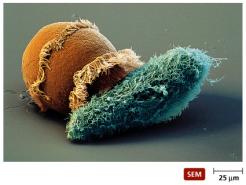
Shorter and more numerous than flagella.
Move cells or substances past the cell surface.
Eukaryotic Cell Walls and Membranes
Fungi, algae, and plants have cell walls made of polysaccharides (cellulose, chitin, agar, etc.).
Animal cells lack cell walls but have ECMs.
Eukaryotic membranes contain cholesterol and other steroids for fluidity and use endocytosis/exocytosis for transport.
Cytoplasm of Eukaryotes
Contains membrane-bound organelles and non-membranous structures (ribosomes, cytoskeleton, centrioles).
Cytoskeleton
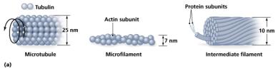
Provides shape, anchors organelles, and enables movement.
Composed of microtubules (tubulin), microfilaments (actin), and intermediate filaments (various proteins).
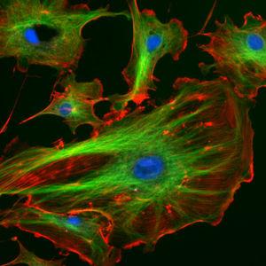
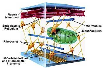
Ribosomes
Eukaryotic ribosomes are 80S (larger than prokaryotic 70S), composed of 60S and 40S subunits.
Sites of protein synthesis (translation).
Nucleus
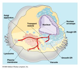
Largest organelle, contains most DNA, surrounded by a double membrane (nuclear envelope) with nuclear pores.
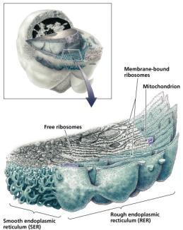
Endomembrane System
Endoplasmic Reticulum (ER): Network of membranes; SER synthesizes lipids, RER (with ribosomes) processes and transports proteins.
Golgi Bodies: Modify, package, and sort proteins and lipids for secretion or delivery to other organelles.
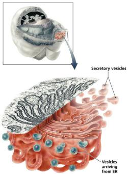
Mitochondria and Chloroplasts
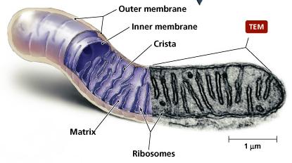
Mitochondria: Double-membraned organelles that produce ATP; contain their own DNA and 70S ribosomes.
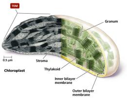
Chloroplasts: Double-membraned, light-harvesting organelles in plants and algae; also contain DNA and 70S ribosomes.
Other Organelles
Lysosomes: Contain digestive enzymes for breaking down waste.
Peroxisomes: Degrade toxic substances.
Vacuoles/Vesicles: Store and transport substances within the cell.
Additional info: Many antibiotics target differences in prokaryotic cell wall and ribosome structure, making these features clinically significant. The "fluid mosaic model" is a unifying concept for all biological membranes, and the "endomembrane system" is crucial for protein and lipid trafficking in eukaryotes.
