 Indietro
Cellular Structure and Functions in Prokaryotes: Morphology, Membranes, Walls, Surface Structures, and Locomotion
Guida di studio - Note intelligenti
 Appunti personalizzati basati sui tuoi materiali, ampliati con definizioni chiave, esempi e contesto.
Cell Shape and Size
Major Cell Morphologies
Bacterial cells exhibit a variety of shapes and arrangements, which are genetically determined and influenced by natural selection. Understanding these morphologies is fundamental to microbiology, as cell shape can affect nutrient uptake, motility, and pathogenicity.
Coccus: Spherical or oval-shaped cells. Variants include Sarcina (3D cubes), Strepto- (chains), and Staphylo- (grape-like clusters).
Rod/Bacilli: Cylindrical cells, often found singly or in chains.
Spirillum: Spiral-shaped, rigid cells.
Spirochete: Flexible, spiral-shaped cells.
Club-shaped: Cells with a wider end.
Filamentous: Long, thread-like cells.
Budding and appendaged bacteria: Cells with stalks or hyphae.
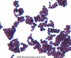
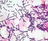
Example: Staphylococcus aureus forms grape-like clusters, while Streptococcus forms chains. 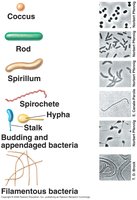
Cell Division Mechanisms
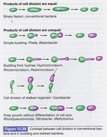
Bacteria can divide by binary fission, budding, or other specialized mechanisms. The products of cell division may be equal or unequal, depending on the species.
Binary fission: Conventional bacteria divide into two equal cells.
Budding: Some bacteria produce unequal daughter cells, such as Hyphomicrobium and Caulobacter.
Cell Size
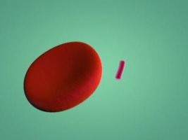
Bacterial cell size ranges from 0.2 µm to over 700 µm in diameter. Most rod-shaped bacteria are 0.5–4.0 µm in diameter and less than 15 µm in length. Eukaryotic cells are generally larger, ranging from 10 to over 200 µm in diameter.
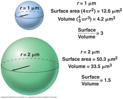
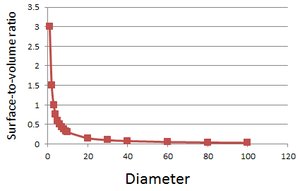
Size matters: Smaller cells have a higher surface-to-volume ratio, which allows for faster nutrient uptake, growth, and adaptation.
Lower size limit: Cells must be large enough to house essential biomolecules; theoretical minimum diameter is about 0.15 µm.
Example: Thiomargarita namibiensis is a giant bacterium, visible to the naked eye. 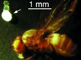
The Cytoplasmic Membrane and Transport
Overall Cellular Structure in Bacteria
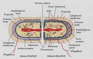
Bacterial cells are surrounded by a cytoplasmic membrane, which is essential for maintaining cellular integrity and mediating transport.
The Cytoplasmic Membrane
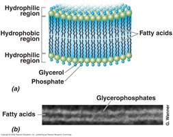
The cytoplasmic membrane is composed of membrane lipids and proteins. It forms a bilayer that provides fluidity and allows molecular interactions.
Phospholipids: Form a bilayer, providing fluidity.
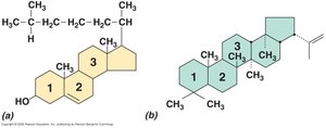
Hopanoids: Rigid planar molecules that stabilize the membrane; sterols are absent in most prokaryotes except methanotrophs and mycoplasmas.
Archaeal Membrane
Archaeal membranes differ from bacterial membranes in their chemical structure.
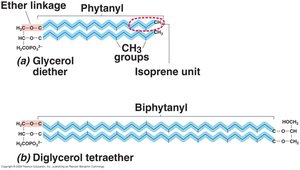
Ether linkage: Archaeal lipids have ether bonds, while bacterial lipids have ester bonds.
No fatty acids: Archaeal lipids use isoprene units.
Lipid monolayer: Hyperthermophiles may have a monolayer for increased stability.
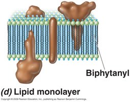
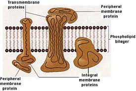
Membrane Proteins
Membrane proteins are crucial for transport, signaling, and structural integrity.
Transmembrane proteins: Span the membrane, often in clusters.
Integral proteins: Embedded within the membrane.
Peripheral proteins: Associated with the membrane surface or other proteins.
Lipoproteins: Have lipid tails, anchoring them to the membrane.
Functions of Cytoplasmic Membranes
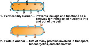
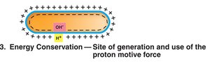
The cytoplasmic membrane serves as a permeability barrier, protein anchor, and site of energy conservation.
Permeability barrier: Prevents diffusion of polar/charged molecules; water can freely diffuse.
Protein anchor: Site for transport and channel proteins.
Energy conservation: Maintains charge separation (proton motive force) for ATP synthesis and other energy-requiring functions.
Membrane Transport Systems
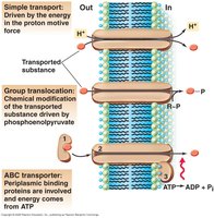
Transport across the cytoplasmic membrane requires specialized proteins and occurs via three main systems.
Simple transport: Driven by proton motive force.
Group translocation: Chemical modification of transported substance, driven by phosphoenolpyruvate.
ABC transporter: Uses ATP hydrolysis and substrate binding proteins.
Transport Events
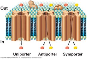
Uniport: Solute moves in one direction (e.g., potassium uniporter).
Antiport: Solute moves in opposite direction to another substance (e.g., sodium-proton antiporter).
Symport: Solute moves together with another substance (e.g., lac permease).
Cell Walls of Prokaryotes
Bacterial Cell Wall
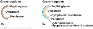
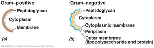
The cell wall provides rigidity and protection. Its structure varies between Gram-positive and Gram-negative bacteria.
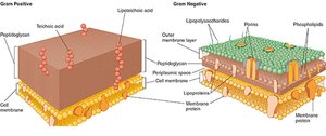
Peptidoglycan (murein): A 3D net-like structure; thick in Gram-positive, thin and sandwiched in Gram-negative.
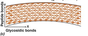
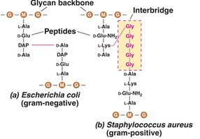
Peptidoglycan Composition
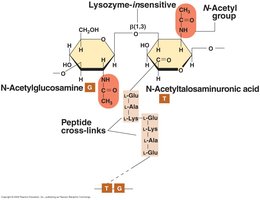
Backbone: Alternating N-acetylglucosamine (G) and N-acetylmuramic acid (M).
Tetrapeptide at M: DAP in Gram-negative, Lys in Gram-positive.
Crosslinkage: Direct in Gram-negative, via Gly interbridge in Gram-positive.
Teichoic acids: Present only in Gram-positive, providing negative charge.
Cell Envelope Structure
Gram-positive bacteria lack a periplasmic space, but recent findings suggest a functionally similar space exists between the cytoplasmic membrane and cell wall.
Outer Membrane: Gram-Negative Bacteria
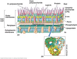
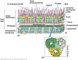
The outer membrane contains lipopolysaccharide (LPS), porin proteins, and forms a barrier to protect the cell.
LPS: Polysaccharide linked to lipid; can act as endotoxin (e.g., Salmonella).
Porin proteins: Channels for small hydrophilic molecules.
Periplasm
Location: Space between outer and cytoplasmic membrane.
Contents: Hydrolytic enzymes, binding proteins, chemoreceptors, Sec proteins.
LPS Structure
O-antigen (O-polysaccharide): Variable region.
Core polysaccharide: Conserved region.
Lipid A: Endotoxic component.
Archaeal Cell Wall
Archaeal cell walls are composed of pseudomurein, which is similar to bacterial peptidoglycan but has distinct chemical differences.
N-acetyltalosaminuronic acid (T): Replaces M of murein.
L-isomer amino acids: Only L-isomers present.
β-1,3 backbone linkage: Lysozyme insensitive.
Surface Appendages and Inclusions
Capsule/Slime Layer, Pili, and Fimbriae
Surface structures play key roles in attachment, protection, and motility.
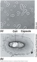
Capsule/slime layer: Outermost polysaccharide layer; capsule is tightly bound, slime layer is loosely bound. Functions include surface attachment, resistance to phagocytosis, and desiccation.
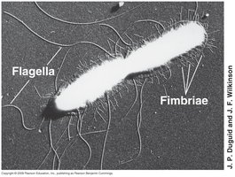
Fimbriae: Filamentous protein structures for attachment.
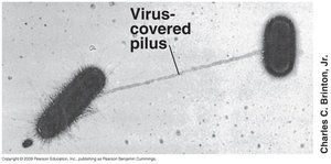
Pili: Longer than fimbriae; involved in genetic exchange (conjugation) and twitching motility.
Inclusion Bodies
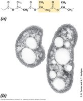
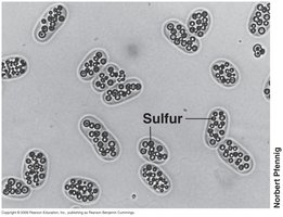
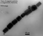
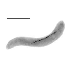
Bacteria store energy and carbon in inclusion bodies, which are often formed in response to stress.
Polyhydroxybutyrate: Energy and carbon storage.
Polyphosphate: Energy storage.
Sulfur granules: Energy storage.
Magnetosomes: Magnetic storage, allowing response to magnetic fields; composed of magnetite (Fe3O4).
Gas Vesicles
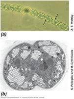
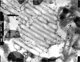
Gas vesicles are hollow, spindle-like protein structures that provide buoyancy, mainly in phototrophic and aquatic bacteria.
Endospores
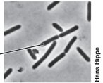
Endospores are differentiated, highly resistant cell structures formed by certain Gram-positive bacteria (e.g., Bacillus and Clostridium) for survival under adverse conditions.
Resistant to: Heat, desiccation, radiation, nutrient depletion.
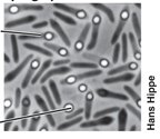
Structure: Exosporium (outer protein layer), spore coat (spore-specific proteins), cortex (loosely crosslinked peptidoglycan), core (cellular essentials).
Life cycle: Sporulation → germination → reproduction.
Microbial Locomotion
Flagella and Motility
Flagella are whip-like appendages that enable motility in bacteria. Their arrangement and mechanism of action vary among species.
Arrangement: Peritrichous (all over), polar (one or both ends), lophotrichous (tufts).
Structure: Basal body (rings + rod), hook, filament.
Rotation: Driven by H+ motor; direction determines movement (run or tumble).
Gliding Motility
Some bacteria move without flagella, using gliding motility, which requires contact with a solid surface and is slower and smoother than flagellar movement.
Twitching motility: Extension and retraction of pili.
Slime extrusion: Excretion of slime for movement.
Motility protein mediated: Driven by membrane proteins and H+ flux.
Microbial Taxis
Bacteria can move in response to environmental stimuli.
Chemotaxis: Movement toward or away from chemical attractants.
Phototaxis: Movement in response to light.
Example: Flagellated bacteria swim toward nutrients or light sources. 

