 Indietro
Direct and Indirect Staining in Microbiology: Principles, Procedures, and Applications
Guida di studio - Note intelligenti
 Appunti personalizzati basati sui tuoi materiali, ampliati con definizioni chiave, esempi e contesto.
Microscopy and Staining in Microbiology
Introduction to Staining and Microscopy
Staining is a fundamental technique in microbiology that enhances the contrast between bacterial cells and their background, allowing for the observation of cell morphology, arrangement, and other cellular structures. Microscopy, particularly bright-field compound light microscopy, is the primary method used to visualize stained bacterial samples.
Contrast: Staining increases contrast, making colorless bacteria visible under the microscope.
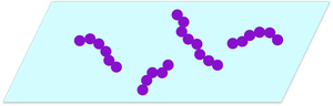
Cell Morphology: Enables identification of cell shapes (cocci, bacilli, spirilla) and arrangements (chains, clusters).
Microscope Components: Key parts include ocular lens, objective lenses, stage, condenser, diaphragm, and focusing knobs.
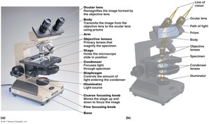
Key Concepts in Microscopy
Wavelength of Radiation: Determines resolution; shorter wavelengths yield higher resolution.
Magnification: Enlargement of the specimen image.
Resolution: Ability to distinguish two close objects as separate.
Contrast: Difference in light intensity between specimen and background.
Chemical Principles of Bacterial Staining
Nature of Stains and Dyes
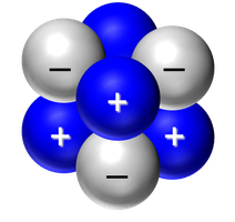
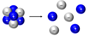
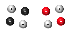
Stains used in microbiology are typically salts composed of positively-charged and negatively-charged ions. The colored ion determines the staining properties.
Salt Structure: Compound of a positive ion and a negative ion.
Example: Methylene blue chloride dissociates into blue (positive) methylene blue ions and colorless (negative) chloride ions.
Types of Dyes
Basic Dyes: Colored portion is the positive ion (e.g., methylene blue, crystal violet, safranin).
Acidic Dyes: Colored portion is the negative ion (e.g., nigrosin, congo red).
Interaction of Dyes with Bacterial Cells
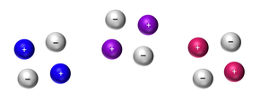
Bacterial cell walls possess a slight negative charge, influencing how basic and acidic dyes interact with the cells.
Basic Dyes: Positively-charged colored ions are attracted to the negatively-charged cell wall, resulting in direct staining of the organism.
Acidic Dyes: Negatively-charged colored ions are repelled by the cell wall, depositing around the organism and resulting in indirect (negative) staining.
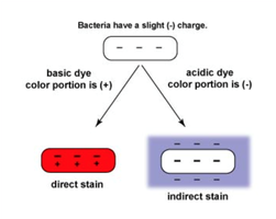
Direct (Basic) and Indirect (Acidic) Staining
Direct Staining (Basic Dye)
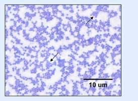
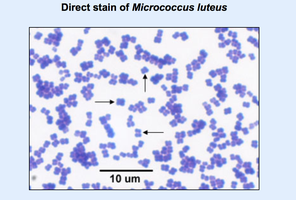
Direct staining uses basic dyes to color the bacterial cells themselves, allowing for clear visualization of cell morphology and arrangement.
Procedure: Fix the sample (heat or chemical), apply basic dye, rinse, and observe under oil immersion.
Examples: Crystal violet, safranin.
Result: Cells appear colored against a clear background.
Indirect Staining (Acidic Dye)
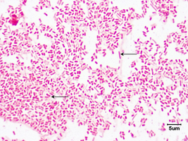
Indirect staining uses acidic dyes to color the background, leaving the bacterial cells unstained and visible as clear areas.
Procedure: Mix bacteria with acidic dye, spread on slide, air dry (no heat fixation), observe under oil immersion.
Examples: Nigrosin, congo red.
Result: Cells appear as clear or white shapes against a dark background.

Staining Procedures
Acidic (Indirect) Staining Procedure
Acidic staining is used to observe cell morphology without heat fixation, preserving cell shape and arrangement.
Place a drop of acidic dye (e.g., nigrosin) on a slide.
Add bacteria and mix gently.
Use a second slide to spread the mixture across the first slide.
Allow to air dry completely; do not heat fix.
Observe under oil immersion microscopy.
Basic (Direct) Staining Procedure
Direct staining involves fixing the bacteria to the slide and applying a basic dye.
Label slide and add a drop of water.
Add bacteria and mix to form a cloudy suspension.
Spread to form a thin film and air dry.
Heat fix by holding near microincinerator for 10 seconds.
Apply basic dye (e.g., crystal violet) for 60 seconds.
Rinse gently and blot dry.
Observe under oil immersion microscopy.
Scientific Principles Behind Staining
Chemical and Physical Principles
Electrostatic Attraction: Basic dyes (positive ions) bind to negatively-charged cell walls; acidic dyes (negative ions) are repelled.
Fixation: Heat or chemical fixation adheres cells to the slide and preserves morphology.
Contrast Enhancement: Staining improves contrast, enabling detailed observation.
Applications and Skills Assessment
Practical Skills
Prepare smears and perform simple stains on bacterial samples.
Focus slides under oil immersion objective and determine cell shape and arrangement.
Identify direct and indirect stained smears.
Comparison of Direct and Indirect Staining
Staining Method | Dye Type | Cell Appearance | Background Appearance | Fixation |
|---|---|---|---|---|
Direct Staining | Basic (positive ion) | Colored | Clear | Heat or chemical |
Indirect Staining | Acidic (negative ion) | Clear/white | Dark/colored | No heat fixation |
Common Questions and Troubleshooting
Purpose of Staining: To improve contrast and enable microscopic observation of cell morphology and arrangement.
Fixing Cells: Adheres cells to slide; achieved by heat or chemical methods.
Stain Types: Basic (direct) and acidic (indirect); examples provided above.
Oil Immersion: Used with 100X objective to increase resolution by reducing light refraction.
Potential Errors: Heat fixing before air drying can distort cell morphology; omitting fixation may result in cells washing off.
Summary Table: Staining Procedures
Step | Direct Staining | Indirect Staining |
|---|---|---|
Sample Preparation | Smear, air dry, heat fix | Mix with dye, spread, air dry |
Dye Application | Basic dye (e.g., crystal violet) | Acidic dye (e.g., nigrosin) |
Observation | Oil immersion microscopy | Oil immersion microscopy |
