Indietro
Disorders Associated with the Immune System: Hypersensitivity, Autoimmunity, Transplantation, Immunodeficiencies, and AIDS
Guida di studio - Note intelligenti
 Appunti personalizzati basati sui tuoi materiali, ampliati con definizioni chiave, esempi e contesto.
Disorders Associated with the Immune System
Hypersensitivity
Hypersensitivity refers to an exaggerated or inappropriate immune response to an antigen, resulting in tissue damage. It occurs when an individual is sensitized by prior exposure to an antigen (allergen). There are four main types of hypersensitivity reactions, each with distinct mechanisms and clinical manifestations.
Type I (Anaphylactic): Immediate reaction mediated by IgE antibodies, leading to mast cell and basophil degranulation and release of mediators such as histamine.
Type II (Cytotoxic): Antibody-mediated destruction of cells, often involving complement activation.
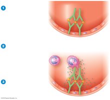
Type III (Immune Complex): Formation of antigen-antibody complexes that deposit in tissues, causing inflammation.
Type IV (Delayed Cell-Mediated): T cell-mediated response, typically delayed, resulting in tissue damage.
Hygiene hypothesis: Suggests that reduced exposure to pathogens may decrease immune tolerance and increase hypersensitivity to harmless antigens.
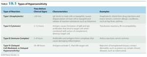
Type of Reaction | Time Before Clinical Signs | Characteristics | Examples |
|---|---|---|---|
Type I (Anaphylactic) | <30 min | IgE binds to mast cells or basophils, causing degranulation and release of histamine and other mediators | Anaphylaxis, allergic reactions (hay fever, asthma) |
Type II (Cytotoxic) | 5–12 hours | Antigen causes formation of IgG and IgM antibodies, which bind to target cell and activate complement, resulting in cell lysis | Transfusion reactions, Rh incompatibility |
Type III (Immune Complex) | 3–8 hours | Antibodies and antigens form complexes that cause inflammation | Arthus reaction, serum sickness |
Type IV (Delayed Cell-Mediated) | 24–48 hours | Antigens activate T cells, which target cells | Allergic contact dermatitis, transplant rejection |
Type I (Anaphylactic) Reactions
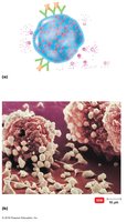
Type I reactions occur within minutes after re-exposure to an allergen. IgE antibodies bind to mast cells and basophils, triggering degranulation and release of mediators such as histamine, leukotrienes, and prostaglandins. These mediators cause increased vascular permeability, smooth muscle contraction, and mucus secretion.
Systemic anaphylaxis: Severe, potentially fatal reaction; treated with epinephrine.
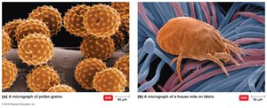
Localized anaphylaxis: Symptoms depend on the route of entry (e.g., hives, hay fever, asthma).
Preventing Anaphylactic Reactions
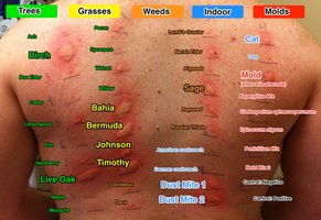
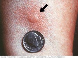
Allergy skin tests involve inoculating antigens beneath the epidermis to detect rapid inflammatory reactions (wheal formation). Desensitization therapy involves repeated injections of increasing doses of antigen to stimulate IgG production, which acts as a blocking antibody to neutralize the allergen before it can trigger IgE-mediated responses.
Blocking antibody: IgG that intercepts and neutralizes antigens.


Type II (Cytotoxic) Reactions
Type II reactions involve IgG or IgM antibodies binding to antigens on cell surfaces, activating complement and causing cell lysis or damage by macrophages. This mechanism is important in blood group incompatibilities and certain drug-induced reactions.
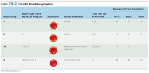
ABO blood group system: Antibodies form against carbohydrate antigens (A, B, or both) on RBCs. Type O RBCs lack these antigens.
Rh blood group system: Rh factor (Rh+) is present in most people. Rh incompatibility can cause hemolytic disease of the newborn (HDNB).
Drug-induced cytotoxic reactions: Drugs can bind to platelets or other cells, forming antigenic complexes that are targeted by antibodies.
Blood Group | Erythrocyte or Red Blood Cell Antigens | Plasma Antibodies | Cells That Can Be Received | Frequency in U.S. Population |
|---|---|---|---|---|
A | A | Anti-B | A, O | White: 41%, Black: 27%, Asian: 28% |
B | B | Anti-A | B, O | White: 10%, Black: 20%, Asian: 27% |
AB | A and B | Neither anti-A nor anti-B | A, B, AB, O | White: 4%, Black: 4%, Asian: 7% |
O | Neither A nor B | Anti-A and anti-B | O | White: 45%, Black: 49%, Asian: 38% |
Type III (Immune Complex) Reactions
Type III reactions occur when antibodies form against soluble antigens, creating immune complexes that deposit in tissues and activate complement, causing inflammation. This is seen in conditions such as glomerulonephritis and serum sickness.
Glomerulonephritis: Immune complexes cause inflammatory damage to kidney glomeruli.
Type IV (Delayed Cell-Mediated) Reactions
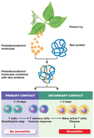
Type IV reactions are mediated by T cells and occur hours to days after exposure to an antigen. Antigens are phagocytized and presented to T cells, causing sensitization. Upon re-exposure, memory T cells release cytokines that cause tissue damage.
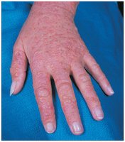
Allergic contact dermatitis: Haptens combine with skin proteins, triggering an immune response (e.g., poison ivy, cosmetics, metals, latex).
Autoimmune Diseases
Mechanisms and Types
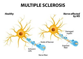
Autoimmune diseases occur when the immune system loses self-tolerance and attacks self-antigens, causing organ damage. Self-tolerance is the ability to distinguish self from nonself. Autoimmune diseases are classified as cytotoxic, immune complex, or cell-mediated.
Cytotoxic: Antibodies react with cell-surface antigens (e.g., multiple sclerosis, Graves' disease, myasthenia gravis).
Immune complex: Immune complexes deposit in tissues (e.g., systemic lupus erythematosus, rheumatoid arthritis).
Cell-mediated: T cells attack tissues (e.g., insulin-dependent diabetes mellitus, psoriasis).
Reactions Related to the Human Leukocyte Antigen (HLA) Complex
HLA Complex and Disease Susceptibility
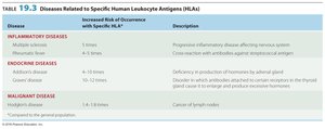
Histocompatibility antigens are self-antigens on cell surfaces encoded by the major histocompatibility complex (MHC). In humans, these genes are called the human leukocyte antigen (HLA) complex. Certain HLAs are associated with increased susceptibility to specific diseases.
Disease | Increased Risk of Occurrence with Specific HLA | Description |
|---|---|---|
Inflammatory diseases | 1.5 times | Progressive inflammatory disease affecting nervous system |
Endocrine diseases | 6–10 times, 40–150 times | Deficiency in production of hormones by adrenal gland; excessive thyroid hormone production |
Malignant disease | 1.5–2 times | Cancer of lymph nodes |
Transplantation and Graft Types
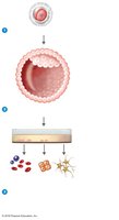
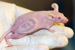
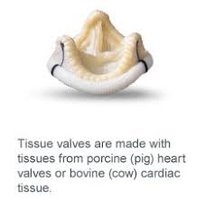
Transplants may be rejected by T cells, macrophages, and complement-fixing antibodies. Tissue typing is essential to match donor and recipient HLAs. Transplants to privileged sites (e.g., cornea, heart valves) are less likely to be rejected. Stem cells, both embryonic and adult, are used to regenerate tissues and organs.
Autograft: Use of one's own tissue.
Isograft: Use of identical twin's tissue.
Allograft: Use of tissue from another person.
Xenotransplantation: Use of nonhuman tissue (e.g., pig heart valves).

The Immune System and Cancer
Immune Response to Cancer
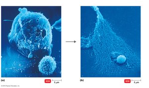
The immune system removes cancer cells through immune surveillance. Cancer cells express tumor-associated antigens, which are recognized by cytotoxic T lymphocytes (CTLs) and macrophages. However, cancer cells may evade immune detection by rapid reproduction, lack of antigenic epitopes, or becoming vascularized.
Immunotherapy for Cancer
Endotoxins: Stimulate TNF to interfere with cancer blood supply.
Vaccines: Used for prophylaxis (e.g., Gardasil for cervical cancer).
Monoclonal antibodies: Target specific tumor antigens (e.g., Herceptin).
Immunotoxins: Combine monoclonal antibodies with toxic agents to selectively kill tumor cells.
Immunodeficiencies
Types and Examples
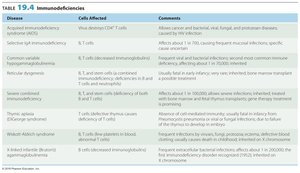
Immunodeficiencies are characterized by insufficient immune responses. They may be congenital (due to genetic defects) or acquired (develop during life due to drugs, cancers, or infections).
Disease | Cells Affected | Comments |
|---|---|---|
Acquired immunodeficiency syndrome (AIDS) | Viral destruction of CD4+ T cells | Allows cancer and bacterial, viral, fungal, and protozoan diseases; usually fatal infection |
Selective IgA immunodeficiency | B cells | Affects about 1 in 700; causes frequent mucosal infections; specific cause unknown |
Common variable immunodeficiency | B cells | Rare; affects adults; increased susceptibility to infections |
Severe combined immunodeficiency | T cells and B cells | Rare; affects about 1 in 100,000; severe infections; treated with bone marrow transplant |
Acquired Immunodeficiency Syndrome (AIDS)
Origin and Structure of HIV
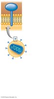
AIDS is caused by the human immunodeficiency virus (HIV), a retrovirus that selectively infects CD4+ T helper cells. HIV originated from cross-species transmission and was first identified in the early 1980s. The virus contains two identical RNA strands, a phospholipid envelope, and glycoprotein spikes (gp120 and gp41).
HIV Infectiveness and Pathogenicity
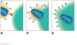
HIV spreads via dendritic cells and macrophages to lymphoid organs, where it infects activated T helper cells. The gp120 spike binds to the CD4 receptor and CCR5 or CXCR4 coreceptors, facilitating viral entry. Once inside, viral RNA is reverse transcribed into DNA and integrated into the host genome.
Latent and Active HIV Infection
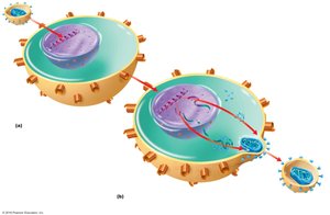
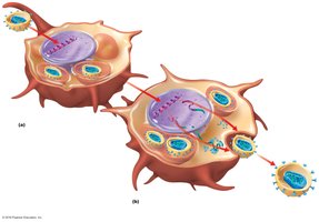
HIV infection can be latent (provirus integrated into host DNA) or active (new viruses bud from the host cell). Latently infected cells serve as reservoirs for HIV, making eradication difficult. The virus undergoes rapid antigenic changes and mutation.
Subtypes and Stages of HIV Infection
HIV-1: Most common, related to chimpanzee viruses.
HIV-2: Less pathogenic, related to monkey viruses.
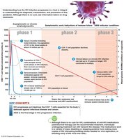
The progression of HIV infection occurs in three phases:
Phase 1: Asymptomatic or lymphadenopathy
Phase 2: Steady decline in CD4+ T cells; persistent infections
Phase 3: AIDS develops; CD4+ count below 200 cells/μl; indicator conditions
HIV Transmission and Global Impact
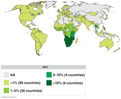
HIV is transmitted via intimate sexual contact, breast milk, transplacental infection, contaminated needles, organ transplants, and blood transfusion. The virus survives for hours outside cells and days inside cells. The global impact is significant, with the highest prevalence in sub-Saharan Africa.
Preventing and Treating AIDS
Prevention includes condom use, discouraging sexual promiscuity, and sterile needles. Treatment involves antiretroviral drugs targeting various stages of the HIV life cycle:
Fusion/entry inhibitors: Block viral entry (e.g., enfuvirtide, maraviroc).
Reverse transcriptase inhibitors: Block viral RNA to DNA conversion (e.g., tenofovir, emtricitabine, efavirenz).
Integrase inhibitors: Prevent integration of viral DNA (e.g., raltegravir).
Protease inhibitors: Prevent maturation of viral proteins (e.g., atazanavir, indinavir, saquinavir).
Maturation inhibitors: Tetherins prevent viral release.
Highly active antiretroviral therapy (HAART) uses drug combinations to minimize resistance.