 Indietro
Endospore Staining and Bacterial Motility: Lab Study Guide
Guida di studio - Note intelligenti
 Appunti personalizzati basati sui tuoi materiali, ampliati con definizioni chiave, esempi e contesto.
Endospore Staining and Bacterial Motility
Introduction
This study guide covers the principles and laboratory techniques for observing bacterial endospores and motility. It includes the biology of endospore formation, staining protocols, and the structural basis of bacterial motility, with emphasis on flagella and their arrangements.
Bacterial Endospores
Definition and Significance
Endospores are highly resistant, dormant structures formed by certain bacteria, primarily Bacillus and Clostridium species.
They enable bacteria to survive extreme conditions such as heat, desiccation, radiation, and chemical disinfectants.
Endospore-forming bacteria include pathogens responsible for diseases like anthrax (Bacillus anthracis), tetanus (Clostridium tetani), and botulism (Clostridium botulinum).
Stages of Endospore Formation (Sporulation)
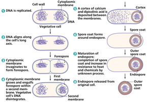
Sporulation is a complex process triggered by harsh environmental conditions. The main stages are:
DNA replication in the vegetative cell.
Alignment of DNA along the cell's long axis.
Invagination of the cytoplasmic membrane to form a forespore.
Engulfment of the forespore by a second membrane; mother cell DNA disintegrates.
Deposition of a cortex of calcium and dipicolinic acid between membranes.
Formation of a spore coat around the endospore.
Maturation of the endospore, increasing resistance to heat and chemicals.
Release of the mature endospore from the degraded mother cell.
Endospore Structure
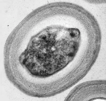
The endospore consists of a core (containing DNA and ribosomes), a cortex (peptidoglycan layer), and a tough spore coat (protein layers).
The spore coat is responsible for the extreme resistance of endospores.
Endospore Germination
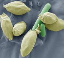
When environmental conditions become favorable, endospores can germinate and return to the vegetative state within approximately 90 minutes.
Endospore Staining
Principle and Importance
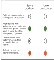
Endospores are difficult to stain due to their tough spore coat; special staining techniques are required.
The primary stain is malachite green, which is forced into the spore by heat.
After staining, water is used as a decolorizer to remove the stain from vegetative cells but not from endospores.
The counterstain is safranin, which stains the vegetative cells pink/red.
Endospore Staining Procedure
Prepare a bacterial smear on a clean slide and allow it to air dry.
Heat-fix the smear by passing it through a flame or using a microincinerator.
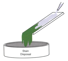
Place blotting paper over the smear and saturate with malachite green.
Steam the slide over boiling water for 5 minutes, adding more stain as needed to prevent drying.
Remove the blotting paper and rinse the slide with water to decolorize the vegetative cells.
Counterstain with safranin for 1 minute, rinse, and blot dry.
Results Interpretation
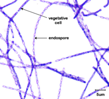
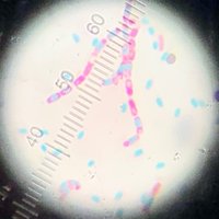
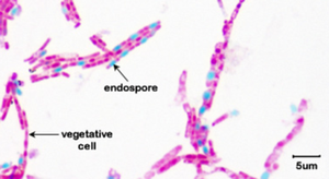
Endospores appear green under the microscope, while vegetative cells appear pink/red.
Bacterial Motility
Definition and Importance
Motility is the ability of bacteria to move independently, usually through liquid environments.
Motility is primarily mediated by flagella, which are whip-like appendages.
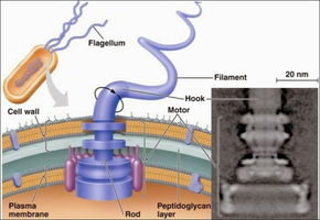
Structure of Bacterial Flagella
Flagella are composed of three main parts:
Filament: The long, helical structure extending from the cell surface.
Hook: A flexible coupling between the filament and the basal body.
Basal body: Anchors the flagellum to the cell wall and plasma membrane; acts as a rotary motor.
Flagellar Arrangements
Monotrichous: Single flagellum at one pole (e.g., Vibrio cholerae).
Amphitrichous: Single flagellum at both poles (e.g., Spirillum species).
Lophotrichous: Two or more flagella at one or both poles (e.g., Spirillum species).
Peritrichous: Flagella distributed over the entire cell surface (e.g., Proteus species).
Mechanism of Motility
Bacteria move by rotating their flagella, which can switch between counterclockwise (smooth swimming) and clockwise (tumbling) rotation.
This movement pattern is called a biased random walk, allowing bacteria to migrate toward attractants (chemotaxis, phototaxis, magnetotaxis).
Axial Filaments
Axial filaments are bundles of endoflagella found in spirochetes (e.g., Borrelia species).
They are located between the cell wall and outer membrane, enabling corkscrew-like movement.
Detecting Bacterial Motility
Microscopy Methods
Phase contrast microscopy: Enhances contrast of live, unstained cells; motile bacteria appear darker than the background.
Dark-field microscopy: Illuminates specimens against a dark background; motile bacteria appear bright.
Flagella Staining
Flagella are too thin to be seen with standard light microscopy; special stains are used to increase their diameter by precipitation.
Motility Test Medium
A semi-solid agar (0.3%) is used to detect motility.
Motile bacteria move away from the stab line, causing diffuse growth throughout the medium.
Non-motile bacteria grow only along the stab line.
Proper inoculation involves stabbing the medium straight down without twisting the loop.
Review Questions and Key Concepts
What are endospores? Name two genera that form endospores.
What is the metabolically active, growing form of the bacterium that forms an endospore called?
Know the different stages of endospore formation.
When do endospores become vegetative cells?
What are the two important conditions required to stain endospores?
What are the different steps of endospore staining?
Name the primary and secondary stains.
What stain do the vegetative cells take up and what stain do the endospores take up?
What is used as a decolorizing agent in endospore staining?
Why do you steam the slide while staining with malachite green?
What color do the vegetative cells and endospores appear after staining?
What is motility? What are the structures involved in bacterial motility called?
What are the three basic parts of a flagellum?
What is chemotaxis? What is biased random walk?
How do you detect motility using motility test medium?
How can you differentiate between motile and non-motile organisms using motility medium?
Summary Table: Endospore Staining Results
Step | Spore Producer | Spore Nonproducer |
|---|---|---|
Cells and spores prior to staining | Transparent | Transparent |
After malachite green + heat | Cells and spores green | Cells green |
Decolorization with water | Cells colorless, spores green | Cells colorless |
Counterstain with safranin | Cells red, spores green | Cells red |
Additional info: The use of 3% agar instead of 0.3% in motility test medium would prevent motile bacteria from moving, as the medium would be too firm.
