 Indietro
Functional Anatomy of Prokaryotic and Eukaryotic Cells: Study Notes
Guida di studio - Note intelligenti
 Appunti personalizzati basati sui tuoi materiali, ampliati con definizioni chiave, esempi e contesto.
Functional Anatomy of Prokaryotic and Eukaryotic Cells
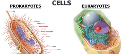
Two Kinds of Cells
Cells are classified into two fundamental types: prokaryotes and eukaryotes. This distinction is foundational in microbiology, as it determines cellular structure, function, and classification of organisms.
Prokaryotes: Include Bacteria and Archaea. No known prokaryotic macroorganisms.
Eukaryotes: Include Animals, Plants, Algae, Fungi, and Protozoa. Both macroorganisms and microorganisms exist in this group.
Prokaryotes | Eukaryotes | |
|---|---|---|
Macroorganisms | None Known | Eukarya: Animals, Plants |
Microorganisms | Archaea, Bacteria | Eukarya: Algae, Fungi, Protozoa |

Cell Structure Comparison
Prokaryotic and eukaryotic cells differ in their internal organization and complexity. Understanding these differences is essential for identifying and studying microorganisms.
Prokaryotes: DNA not enclosed in a nuclear membrane, usually a single circular chromosome, no membrane-bound organelles, complex cell wall if present, divide by binary fission, smaller ribosomes, no cytoskeleton.
Eukaryotes: DNA enclosed in a nuclear membrane, multiple chromosomes, associated with histones and non-histones, membrane-bound organelles (e.g., Golgi complex, mitochondria), simple cell wall if present, divide by mitosis, larger ribosomes, cytoskeleton present.
Feature | Prokaryotes | Eukaryotes |
|---|---|---|
DNA Location | Not enclosed in nuclear membrane | Enclosed in nuclear membrane |
Chromosomes | One circular | Multiple, linear |
Histones | Absent | Present |
Organelles | Absent | Present |
Cell Wall | Complex (if present) | Simple (if present) |
Division | Binary fission | Mitosis |
Size | 0.2–20 μm | 10–100 μm |
Ribosomes | Smaller | Larger |
Reproduction | Asexual (may transfer DNA fragments) | Sexual (meiosis) |
Origin of Terms
The terms prokaryote and eukaryote are derived from Greek:
Prokaryote: "prenucleus"
Eukaryote: "true nucleus"
The Prokaryotes
Prokaryotes include Bacteria and Archaea. They are distinguished by their cellular structure and metabolic diversity.
Some bacteria are photosynthetic (e.g., Cyanobacteria).
Species are differentiated by:
Morphology (shape)
Chemical composition (detected by staining)
Nutritional requirements
Biochemical activities
Source of energy (sunlight or chemicals)
Basic Shapes of Bacteria
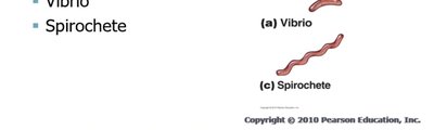
Bacteria exhibit a variety of shapes, which are important for identification and classification.
Bacillus: Rod-shaped
Coccus: Spherical
Spiral: Includes Spirillum, Vibrio, and Spirochete
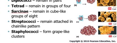
Shapes of Bacterial Cells: Coccus
Cocci are round, spherical, oval, or elongated. Their arrangements are key to identification.
Single
Diplococci: Pairs
Tetrad: Groups of four
Sarcinae: Cube-like groups of eight
Streptococci: Chain-like pattern
Staphylococci: Grape-like clusters
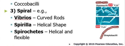
Shapes of Bacterial Cells: Bacillus and Spiral
Bacilli are rod-shaped, and spiral bacteria have distinctive helical forms.
Bacillus: Single, diplobacilli, streptobacilli, coccobacilli
Spiral:
Vibrios: Curved rods
Spirilla: Helical shape
Spirochetes: Helical and flexible
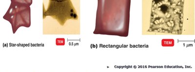
Unusual Bacterial Shapes
Some bacteria exhibit unusual shapes, such as star-shaped (Stella) and square (Haloarcula).
Monomorphic and Pleomorphic Bacteria
Bacterial shape is determined by heredity. Most bacteria are monomorphic (maintain a single shape), but some are pleomorphic (can have more than one genetically controlled shape).
Monomorphic: Environmental factors may change shape, complicating identification.
Pleomorphic: Examples include Rhizobium and Corynebacterium.
Prokaryotic Cell Diagram
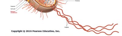
The structure of a typical prokaryotic cell includes the plasma membrane, cell wall, capsule, cytoplasm, ribosomes, nucleoid, and flagella.
Structures External to the Cell Wall: Glycocalyx
The glycocalyx is a sugar coat on the surface of many cells, providing protection and aiding in attachment.
Bacterial glycocalyx is viscous (sticky).
Gelatinous polymer of polysaccharide and/or polypeptide.
Made inside and secreted to the cell surface.
Called capsule if organized and firmly attached; slime layer if unorganized and loose.
Structures External to the Cell Wall: Capsule
The capsule is an important virulence factor for pathogenic bacteria.
Provides protection from phagocytosis (e.g., Bacillus anthracis).
Allows attachment to surfaces (e.g., Klebsiella colonization).
Can serve as a source of nutrition when energy stores are low.
Protects against dehydration.
Capsules are antigenic (contain antigens).

Structures External to the Cell Wall: Flagella
Flagella are long filamentous appendages that propel bacteria, contributing to motility and classification.
Flagellar protein (H antigen) is used to distinguish among serovars.
Four arrangements:
Monotrichous: Single polar flagellum
Amphitrichous: Tuft of flagella on both ends
Lophotrichous: Two or more flagella at one pole
Peritrichous: Flagella distributed over entire cell

Structure of a Prokaryotic Flagellum
A flagellum consists of three basic parts: filament, hook, and basal body.
Filament: Long, outermost region; composed of flagellin protein arranged in intertwining chains forming a helix around a hollow core.
Hook: Filament attached to it; wider than filament; composed of different protein.
Basal body: Anchors flagellum to cell wall and plasma membrane (not shown in the extracted text but standard academic context).
Additional info: The basal body is a critical component for flagellar function, anchoring the flagellum and enabling rotation for motility.
