 Indietro
Functional Anatomy of Prokaryotic and Eukaryotic Cells: Structure, Function, and Comparison
Guida di studio - Note intelligenti
 Appunti personalizzati basati sui tuoi materiali, ampliati con definizioni chiave, esempi e contesto.
Comparing Prokaryotic and Eukaryotic Cells
Overview of Cell Types
Prokaryotic and eukaryotic cells represent two fundamental types of cellular organization in biology. Understanding their structural and functional differences is essential for microbiology.
Prokaryotes: Cells lacking a membrane-bound nucleus; include Bacteria and Archaea.
Eukaryotes: Cells with a true nucleus and membrane-bound organelles; include plants, animals, fungi, and protists.
Key Differences: Chromosome structure, presence of histones, organelles, cell wall composition, and division mechanisms.
Example: Bacteria (prokaryotes) have peptidoglycan cell walls, while plant cells (eukaryotes) have cellulose cell walls.
The Size, Shape, and Arrangement of Bacterial Cells
Bacterial Morphology
Bacteria exhibit a variety of shapes and arrangements, which are important for identification and classification.
Average Size: 0.2–2.0 μm diameter, 2–8 μm length.
Monomorphic: Most bacteria have a single, consistent shape.
Pleomorphic: Some bacteria can vary in shape.
Basic Shapes: Bacillus (rod-shaped), Coccus (spherical), Spiral (vibrio, spirillum, spirochete), Star-shaped, Rectangular.
Example: Bacillus species are rod-shaped, while Staphylococcus forms clusters of spherical cells.
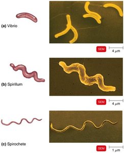
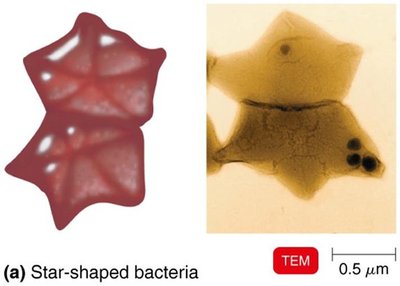
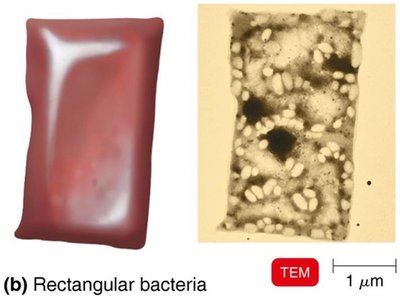
Arrangement of Cocci and Bacilli
Bacterial cells can be arranged in characteristic patterns based on their division and grouping.
Cocci: Diplococci (pairs), Streptococci (chains), Tetrads (groups of four), Sarcinae (cubelike groups of eight), Staphylococci (clusters).
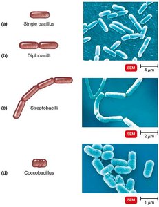
Bacilli: Single bacillus, Diplobacilli (pairs), Streptobacilli (chains), Coccobacillus (short rods).
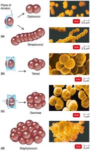
Structure of a Prokaryotic Cell
Cell Components
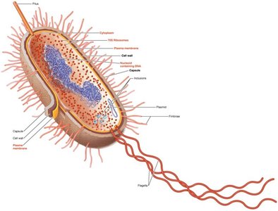
Prokaryotic cells contain several essential structures, each with specific functions.
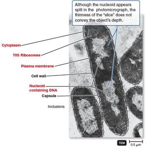
Capsule: Protective layer outside the cell wall.
Cell Wall: Provides structural support and shape.
Plasma Membrane: Controls entry and exit of substances.
Cytoplasm: Site of metabolic activity.
Nucleoid: Region containing the bacterial chromosome (DNA).
Plasmid: Small, circular DNA molecules with non-essential genes.
Ribosomes: Sites of protein synthesis (70S type).
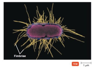
Fimbriae: Hairlike appendages for attachment.
Pili: Involved in motility and DNA transfer.
Flagella: Motility structures.
Inclusions: Reserve deposits of nutrients.
Glycocalyx
Structure and Function
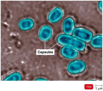
The glycocalyx is an external, viscous, gelatinous layer composed of polysaccharides and/or polypeptides.
Capsule: Neatly organized and firmly attached.
Slime Layer: Unorganized and loose.
Functions: Contributes to virulence, prevents phagocytosis, helps microbes adhere to surfaces, forms biofilms.
Example: Capsules in Bacillus anthracis and Streptococcus pneumoniae enhance pathogenicity.
Flagella, Axial Filaments, Fimbriae, and Pili
Flagella
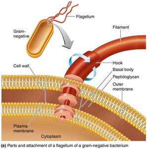
Flagella are filamentous appendages that propel bacteria. They consist of three parts: filament, hook, and basal body.
Filament: Outermost region, composed of flagellin protein.
Hook: Attaches filament to basal body.
Basal Body: Anchors flagellum to cell wall and membrane.
Function: Motility, taxis (movement toward/away from stimuli), identification via H antigens.
Flagellar Arrangements
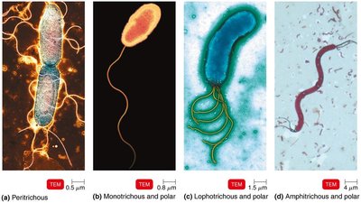
Bacteria exhibit various flagellar arrangements, which aid in identification.
Peritrichous: Flagella distributed over entire cell.
Monotrichous: Single flagellum at one end.
Lophotrichous: Multiple flagella at one end.
Amphitrichous: Flagella at both ends.
Flagellar Movement
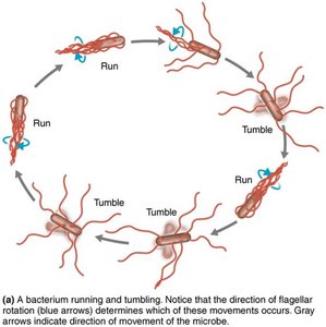
Flagella rotate to produce "runs" and "tumbles," enabling directional movement.
Run: Flagella rotate together for straight movement.
Tumble: Flagella splay outward, causing random movement.
Axial Filaments (Endoflagella)
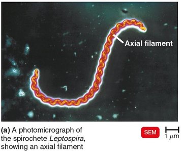
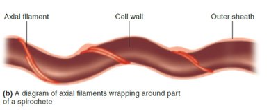
Axial filaments are found in spirochetes and enable corkscrew-like movement.
Structure: Anchored at one end, wrap around cell beneath outer sheath.
Function: Rotation causes cell to move through viscous environments.
Fimbriae and Pili
Fimbriae are hairlike appendages for attachment, while pili are involved in motility and DNA transfer.
Fimbriae: Enable attachment to surfaces and formation of biofilms.
Pili: Facilitate gliding/twitching motility and conjugation (DNA transfer).
The Cell Wall
Structure and Function
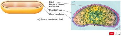
The bacterial cell wall prevents osmotic lysis, protects the cell membrane, and contributes to pathogenicity. It is a major target for antibiotics.
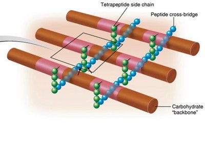
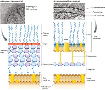
Peptidoglycan: Polymer of repeating disaccharides (N-acetylglucosamine and N-acetylmuramic acid) linked by polypeptides.
Lattice Structure: Provides rigidity and protection.
Penicillin: Interferes with peptide cross-bridges, weakening the cell wall.
Gram-Positive Cell Walls
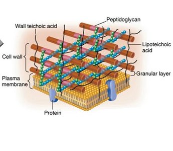
Gram-positive bacteria have thick peptidoglycan layers and teichoic acids.
Teichoic Acids: Link cell wall to plasma membrane, regulate cation movement, provide antigenic specificity.
High Susceptibility: More susceptible to penicillin and lysozyme.
Gram-Negative Cell Walls
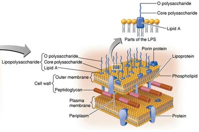
Gram-negative bacteria have thin peptidoglycan, an outer membrane, and a periplasmic space.
Outer Membrane: Contains lipopolysaccharide (LPS), lipoproteins, and phospholipids.
LPS: O polysaccharide (antigen), Lipid A (endotoxin).
Porins: Proteins forming channels.
Low Susceptibility: Less susceptible to penicillin.
Gram Stain Mechanism
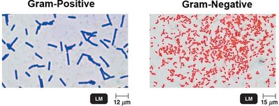
The Gram stain differentiates bacteria based on cell wall structure.
Gram-Positive: Alcohol dehydrates peptidoglycan; crystal violet-iodine complex remains.
Gram-Negative: Alcohol dissolves outer membrane; crystal violet-iodine complex washes out; safranin stains cells.
Atypical Cell Walls
Some bacteria have atypical cell walls, such as acid-fast bacteria and mycoplasmas.
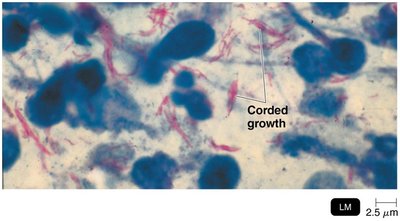
Acid-Fast: Thick peptidoglycan, waxy mycolic acid, stain with carbolfuchsin.
Mycoplasmas: Lack cell walls, have sterols in plasma membrane.
Archaea: Wall-less or walls of pseudomurein (lack NAM and D-amino acids).
The Plasma (Cytoplasmic) Membrane
Structure
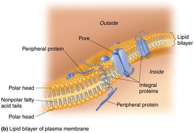
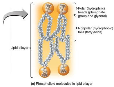
The plasma membrane is a phospholipid bilayer with embedded proteins, forming a fluid mosaic model.
Phospholipid Bilayer: Hydrophilic heads face outward, hydrophobic tails inward.
Proteins: Peripheral, integral, and transmembrane proteins.
Carbohydrates: Glycoproteins and glycolipids for cell recognition.
Function
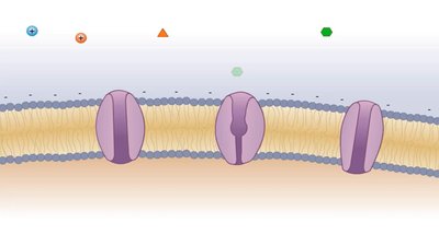
The plasma membrane regulates selective permeability, contains enzymes for ATP production, and may have photosynthetic pigments (chromatophores).
Movement of Materials Across Membranes
Passive Processes
Passive transport does not require energy and includes simple diffusion, facilitated diffusion, and osmosis.
Simple Diffusion: Movement from high to low concentration.
Facilitated Diffusion: Uses transporter proteins for ions and larger molecules.
Osmosis: Net movement of water across a selectively permeable membrane.
Osmotic Solutions
Cells respond differently to isotonic, hypotonic, and hypertonic solutions.
Isotonic: Equal solute concentrations; no net water movement.
Hypotonic: Lower solute outside; water enters cell.
Hypertonic: Higher solute outside; water leaves cell.
Active Processes
Active transport requires energy (ATP) and moves substances against the concentration gradient. Group translocation alters substances as they cross the membrane.
Cytoplasm, Nucleoid, Ribosomes, and Inclusions
Cytoplasm
The cytoplasm is a thick, aqueous, semitransparent substance inside the plasma membrane, containing DNA, ribosomes, and inclusions.
Nucleoid
The nucleoid contains the bacterial chromosome (circular, double-stranded DNA) and plasmids.
Ribosomes
Prokaryotic ribosomes (70S) are the sites of protein synthesis and are targets for specific antibiotics.
Inclusions
Inclusions are reserve deposits of nutrients, such as phosphate, polysaccharides, lipids, sulfur, gas vacuoles, and magnetosomes.
Endospores
Formation and Function
Endospores are highly resistant, dormant structures formed by certain bacteria under nutrient depletion.
Resistant to: Desiccation, heat, chemicals, radiation.
Produced by: Bacillus and Clostridium species.
Sporulation: Endospore formation process.
Germination: Return to vegetative state.
Comparing Eukaryotic and Prokaryotic Structures
Flagella and Cilia
Eukaryotic flagella and cilia are projections used for locomotion or moving substances along the cell surface. They consist of microtubules arranged in a 9+2 array and move in a wavelike manner.
Cell Wall and Glycocalyx
Eukaryotic cell walls are found in plants, algae, and fungi, composed of carbohydrates. The glycocalyx is found in animal cells and strengthens the cell surface, aids in attachment, and cell-cell recognition.
Plasma Membrane
Eukaryotic plasma membranes are similar to prokaryotic membranes but contain sterols and carbohydrates for attachment and recognition. They also perform endocytosis (phagocytosis, pinocytosis, receptor-mediated).
Cytoplasm
Eukaryotic cytoplasm contains a cytoskeleton (microfilaments, intermediate filaments, microtubules) and exhibits cytoplasmic streaming.
Ribosomes
Eukaryotic ribosomes are 80S (60S + 40S subunits), found free in cytoplasm or bound to endoplasmic reticulum. 70S ribosomes are present in mitochondria and chloroplasts.
The Endosymbiotic Theory of Eukaryotic Evolution
Origin and Evidence
The endosymbiotic theory explains the origin of eukaryotes as a result of larger bacterial cells engulfing smaller ones, leading to the development of mitochondria and chloroplasts.
Mitochondria and Chloroplasts: Resemble bacteria in size and shape, have circular DNA, reproduce independently, have bacterial-type ribosomes, and double membranes.
Genomic Evidence: Sequenced genomes are similar to cyanobacteria (chloroplasts) and rickettsia (mitochondria).
Additional info: Eukaryotes branch off from Archaea, supporting the evolutionary relationship.
----------------------------------------
