 Indietro
Functional Anatomy of Prokaryotic and Eukaryotic Cells
Guida di studio - Note intelligenti
 Appunti personalizzati basati sui tuoi materiali, ampliati con definizioni chiave, esempi e contesto.
Prokaryotic Cell Structures
Overview of Prokaryotic Cells
Prokaryotic cells are typically measured in micrometers and are generally longer than they are wide. They lack a nucleus and membrane-bound organelles, distinguishing them from eukaryotic cells.
Bacterial Shapes and Arrangements: Bacteria exhibit various shapes (cocci, bacilli, spirilla) and arrangements (chains, clusters).
Flagella and Glycocalyx: Flagella are used for motility, while the glycocalyx is a protective, adhesive layer outside the cell wall.
Axial Filaments (Endoflagella)
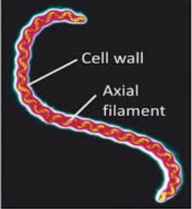
Axial filaments are specialized structures found in spirochetes, appearing as a "rope" twined around the outer surface of the bacteria. They facilitate movement in viscous environments.
Clinical Importance: Spirochetes such as Treponema pallidum (syphilis) and Borrelia burgdorferi (Lyme disease) possess axial filaments.
Fimbriae and Pili
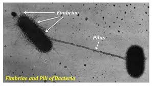
Fimbriae are hairlike structures on the external cell surface, present in few or many numbers, and function primarily in adherence and biofilm formation. Pili are longer, usually one or two per cell, and are involved in twitching motility and conjugation (horizontal DNA transfer).
Fimbriae: Important for adherence; clinical relevance includes Neisseria gonorrhoeae and Escherichia coli.
Pili: Made of pilin protein; main function is conjugation, acting as a bridge for DNA transfer between cells.
Conjugation: Horizontal gene transfer via pilus; vertical transfer is from parent to offspring.
Cell Wall Structure and Function
The cell wall is a complex, semi-rigid structure surrounding the plasma membrane, composed mainly of peptidoglycan (murein). It provides structural support, shape, protection, and prevents cell lysis in hypotonic environments.
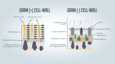
Gram-Positive vs. Gram-Negative: Gram-positive bacteria have a thick peptidoglycan layer with teichoic acids; Gram-negative bacteria have a thin peptidoglycan layer and an outer membrane lacking teichoic acids.
Clinical Importance: Cell wall composition affects pathogenicity and is a target for antibiotics.
Gram-Positive Cell Wall
Thick peptidoglycan layer
Contains teichoic and lipoteichoic acids (move cations, cell growth, antigenic specificity)
Gram-Negative Cell Wall
Thin peptidoglycan layer
Outer membrane with lipoproteins, phospholipids, and lipopolysaccharides (LPS)
Porins for nutrient uptake
Effective barrier to antibiotics and enzymes
Plasma Membrane
The plasma membrane is a phospholipid bilayer with embedded proteins and cholesterol, described by the Fluid Mosaic Model.
Functions: Semi-permeable barrier, site of nutrient breakdown and ATP production, cell signaling.
Transport Mechanisms: Simple diffusion, osmosis, facilitated diffusion, active transport.
Cytoplasm and Internal Structures
The cytoplasm is a thick, aqueous solution containing water, proteins, carbohydrates, lipids, and ions, supported by a cytoskeleton.
Nucleoid Region: Contains bacterial chromosome and plasmids (extra-chromosomal DNA for antibiotic resistance, toxin production).
Ribosomes: Site of protein synthesis; composed of 30S and 50S subunits forming 70S ribosomes.
Inclusions: Reserve deposits for nutrients and metabolic functions.
Types of Inclusions
Metachromatic granules: Inorganic phosphate reserves
Polysaccharide granules: Glycogen and starch reserves
Lipid inclusions: Energy reserves
Sulfur granules: Energy reserves
Carboxysomes: RuBisCO enzymes for CO2 fixation
Gas vacuoles: Buoyancy control
Magnetosomes: Iron oxide inclusions for H2O2 destruction
Eukaryotic Cell Structures
Overview of Eukaryotic Cells
Eukaryotic cells are larger than prokaryotic cells, possess a nucleus, and contain membrane-bound organelles.
Flagella: Few, long, anchored via basal body, made of microtubules.
Cilia: Many, short, anchored via basal body, made of microtubules.
Cell Wall and Glycocalyx
Cell Wall: Found in plants, algae, fungi; composed of carbohydrates (cellulose, chitin, glucan, mannan).
Glycocalyx: External to plasma membrane in animal cells; composed of carbohydrates, glycoproteins, glycolipids; strengthens cell surface and connects cells.
Plasma Membrane
Similar to prokaryotes but with different proteins, carbohydrates, and sterols.
Endocytosis: Includes phagocytosis for nutrient uptake.
Cytoplasm and Cytoskeleton
Cytosol: Fluid portion of cytoplasm.
Cytoskeleton: Network of fibers for structure, cell division, and movement.
Ribosomes
Protein synthesis; composed of 60S and 40S subunits forming 80S ribosomes.
Mitochondria and chloroplasts contain 70S ribosomes (similar to prokaryotes).
Nucleus
Contains DNA instructions; directs protein synthesis.
Chromatin: Nuclear DNA + proteins (histone and non-histone).
Nuclear envelope: Double membrane surrounding nucleus.
Endoplasmic Reticulum (ER)
Rough ER: Ribosomes attached; synthesizes proteins and phospholipids.
Smooth ER: No ribosomes; synthesizes lipids, carbohydrates, cholesterol; detoxification.
Golgi Apparatus
Finishes, sorts, modifies, and ships proteins.
Composed of non-continuous cisterns.
Lysosomes and Vacuoles
Lysosomes: Contain digestive enzymes for macromolecule breakdown.
Vacuoles: Derived from Golgi; primarily in plant cells; regulate water balance.
Mitochondria
Site of ATP synthesis (cellular respiration); double membrane with cristae and matrix.
Bacteria lack mitochondria; cellular respiration occurs in plasma membrane.
Peroxisomes
Contain enzymes for oxidation of organic compounds and detoxification (e.g., hydrogen peroxide).
Selective Toxicity
Drugs that target specific microbes or organelles without harming host cells.
Additional info: Academic context was added to clarify cell structure functions, clinical relevance, and comparisons between prokaryotic and eukaryotic cells.
