 Indietro
Functional Anatomy of Prokaryotic and Eukaryotic Cells: Study Notes
Guida di studio - Note intelligenti
 Appunti personalizzati basati sui tuoi materiali, ampliati con definizioni chiave, esempi e contesto.
Prokaryotic and Eukaryotic Cells
Overview and Definitions
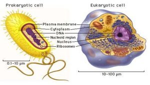
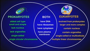
Prokaryotic and eukaryotic cells represent the two fundamental types of cellular organization in biology. Understanding their structural and functional differences is essential for microbiology.
Prokaryotes: Cells lacking a true nucleus and membrane-bound organelles. Includes Bacteria and Archaea.
Eukaryotes: Cells with a true nucleus and membrane-bound organelles. Includes Animals, Plants, Fungi, and Protists.
Structural Comparison
Prokaryotes: One circular chromosome (not in a membrane), no histones, no organelles, cell wall (peptidoglycan in bacteria, pseudomurein in archaea), binary fission.
Eukaryotes: Paired, linear chromosomes (in nuclear membrane), histones, organelles, polysaccharide cell walls, mitotic spindle.
Bacterial Morphology
Basic Shapes of Bacteria
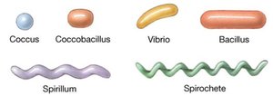
Bacteria exhibit a variety of shapes, which are important for identification and classification.
Bacillus: Rod-shaped
Coccus: Spherical
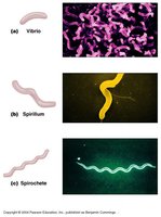
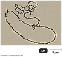
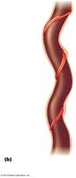
Spiral: Includes vibrio (curved), spirillum (helical, rigid), and spirochete (helical, flexible)
Coccobacillus: Oval, intermediate between cocci and bacilli
Unusual Shapes and Arrangements
Some bacteria are monomorphic (single shape), others are pleomorphic (variable shape).
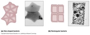
Unusual shapes: star-shaped (Stella), square (Haloarcula).
Arrangement of Bacterial Cells
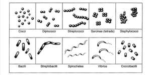
Bacterial cells can be arranged in distinctive patterns, aiding identification.
Cocci: Diplococci (pairs), streptococci (chains), tetrads (groups of four), sarcinae (cubelike), staphylococci (clusters)
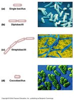
Bacilli: Single, diplobacilli (pairs), streptobacilli (chains), coccobacilli (oval)
Spirals: Vibrios (curved), spirillum (helical with flagella), spirochetes (helical with axial filaments)
Structures External to the Cell Wall
Glycocalyx
The glycocalyx is a sticky, protective layer outside the cell wall, important for adherence and virulence.
Capsule: Neatly organized, protects against phagocytosis.
Slime layer: Unorganized and loose, aids in biofilm formation.
Extracellular polysaccharide (EPS): Facilitates attachment to surfaces.
Flagella, Axial Filaments, Fimbriae, and Pili
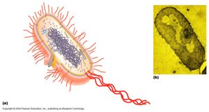
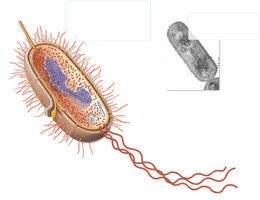
These structures contribute to motility, adherence, and genetic exchange.
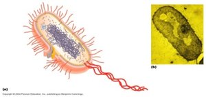
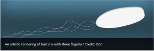
Flagella: Long, whip-like appendages for motility; made of flagellin, anchored by basal body.
Axial filaments: Endoflagella in spirochetes, cause corkscrew motion.
Fimbriae: Short, numerous, hairlike structures for attachment.
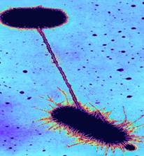
Pili: Longer than fimbriae, used for attachment and DNA transfer (conjugation).
The Cell Wall
Structure and Function
The cell wall is a semi-rigid structure that maintains cell shape and prevents osmotic lysis.
Peptidoglycan: Unique to bacteria, composed of N-acetylglucosamine (NAG) and N-acetylmuramic acid (NAM) linked by polypeptides.
Provides structural integrity and is a target for antibiotics (e.g., penicillin).
Gram-Positive vs. Gram-Negative Cell Walls
Gram-Positive: Thick peptidoglycan, teichoic acids, no outer membrane.
Gram-Negative: Thin peptidoglycan, outer membrane with lipopolysaccharides (LPS), periplasmic space.
Gram Staining Mechanism
Gram staining differentiates bacteria based on cell wall structure.
Gram-Positive: Retain crystal violet stain due to thick peptidoglycan.
Gram-Negative: Lose crystal violet stain, take up counterstain (safranin).
Atypical Cell Walls
Mycoplasmas: Lack cell walls, have sterols in plasma membrane.
Archaea: Wall-less or walls of pseudomurein (lack NAM and D amino acids).
Acid-Fast Cells: High concentration of mycolic acid (waxy), stain better by acid-fast method.
Damage to Cell Walls
Lysozyme: Digests peptidoglycan.
Penicillin: Inhibits peptide bridges in peptidoglycan.
Protoplast: Wall-less cell.
Spheroplast: Wall-less Gram(-) cell.
L form: Wall-less cells that swell into irregular shapes.
Endospores
Formation and Function
Endospores are highly resistant, dormant structures formed by some Gram-positive bacteria for survival under adverse conditions.
Sporulation: Process of endospore formation, not reproduction.
Germination: Return to vegetative state.
Resistant to: Desiccation, heat, chemicals.
Examples: Bacillus (aerobic), Clostridium (anaerobic).
Summary Table: Cell Wall Types
Type | Main Features | Example Organisms |
|---|---|---|
Gram-Positive | Thick peptidoglycan, teichoic acids, no outer membrane | Bacillus, Streptococcus |
Gram-Negative | Thin peptidoglycan, outer membrane with LPS, periplasmic space | Escherichia coli, Pseudomonas |
Acid-Fast | Waxy mycolic acid, weakly Gram-positive | Mycobacterium tuberculosis |
Mycoplasma | No cell wall, sterols in membrane | Mycoplasma pneumoniae |
Archaea | Pseudomurein or wall-less | Halobacterium |
Check Your Understanding
What is the main feature that distinguishes prokaryotes from eukaryotes?
How would you identify streptococci through a microscope?
Why are bacterial capsules medically important?
How do bacteria move?
Why are drugs that target cell wall synthesis useful?
Why are mycoplasmas resistant to antibiotics that interfere with cell wall synthesis?
How do protoplasts differ from L forms?
Describe the functions of endospores, sporulation, and endospore germination.
Under what conditions do endospores form?
