 Indietro
Fundamentals of Microbial Growth and Decontamination: Study Notes
Guida di studio - Note intelligenti
 Appunti personalizzati basati sui tuoi materiali, ampliati con definizioni chiave, esempi e contesto.
Fundamentals of Microbial Growth and Decontamination
Microbial Growth Basics
Microbial growth refers to the increase in the number of cells in a population, primarily through cell division. Understanding microbial growth is essential for laboratory studies, clinical diagnostics, and industrial applications.
Laboratory vs. Natural Growth: In the lab, microbes are often grown as pure cultures, while in nature, they exist in complex communities, often forming biofilms.
Biofilms: Structured communities of microbes attached to surfaces, significant in healthcare due to their resistance to treatment and role in persistent infections.
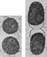
Cell Division Mechanisms: Most bacteria divide by binary fission, but some use budding or spore formation.
Generation Time: The time required for a cell to divide; varies by species and conditions (e.g., E. coli ~20 min, Mycobacterium tuberculosis 15–20 hours).
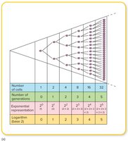
Exponential Growth and Population Doubling
Bacterial populations grow exponentially under favorable conditions, doubling each generation. The number of cells after n generations is given by .
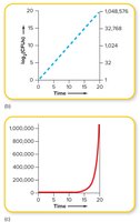
Exponential Growth: Population size increases rapidly; best visualized on a logarithmic scale.
Arithmetic vs. Logarithmic Plots: Logarithmic plots yield straight lines, indicating exponential growth.
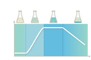
Bacterial Growth Curve in Batch Culture
When bacteria are cultured in a closed system, they exhibit four distinct growth phases:
Lag Phase: Cells adapt to new environment; little division occurs.
Log (Exponential) Phase: Rapid cell division and population growth.
Stationary Phase: Growth rate slows as resources deplete; cell division equals cell death.
Death Phase: Cells die due to waste accumulation and nutrient exhaustion.
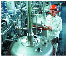
Industrial Application: Chemostats are used to maintain cultures in a specific growth phase for continuous production.
Prokaryotic Growth Requirements
Temperature Requirements
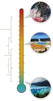
Microbes are classified by their optimal temperature ranges:
Psychrophiles: -20°C to 10°C
Psychrotrophs: 0°C to 30°C (associated with food spoilage)
Mesophiles: 10°C to 50°C (most pathogens)
Thermophiles: 40°C to 75°C
Extreme Thermophiles: 65°C to 120°C
pH and Salinity Requirements
Acidophiles: Grow at pH 1–5 (e.g., Lactobacillus in the vagina)
Neutralophiles: pH 5–8 (most microbes)
Alkaliphiles: pH 9–11 (e.g., in soda lakes)
Halophiles: Thrive in high-salt environments (up to 35%)
Facultative Halophiles: Tolerate higher salt but do not require it (e.g., Staphylococcus aureus)
Oxygen Requirements
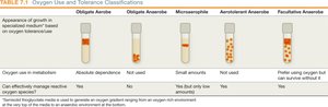
Microbes are classified by their oxygen use and tolerance:
Obligate Aerobes: Require oxygen for metabolism.
Obligate Anaerobes: Cannot tolerate oxygen; lack enzymes to detoxify ROS.
Facultative Anaerobes: Can use oxygen but also grow without it.
Microaerophiles: Require small amounts of oxygen.
Aerotolerant Anaerobes: Tolerate oxygen but do not use it.
Type | Oxygen Use | Growth Pattern |
|---|---|---|
Obligate Aerobe | Yes | Top of tube |
Obligate Anaerobe | No | Bottom of tube |
Facultative Anaerobe | Yes/No | Throughout, more at top |
Microaerophile | Low O2 | Just below surface |
Aerotolerant Anaerobe | No | Evenly throughout |
Microbial Nutrition and Energy Sources
Essential Nutrients and Growth Factors
Macronutrients: Required in large amounts (C, H, O, N, S, P, K, etc.)
Micronutrients: Required in trace amounts (e.g., Fe, Zn, Cu)
Growth Factors: Organic compounds a microbe cannot synthesize and must obtain from the environment (e.g., vitamins, amino acids)
Fastidious Organisms: Require multiple growth factors and complex media for cultivation.
Energy and Carbon Sources
Phototrophs: Use light as an energy source.
Chemotrophs: Obtain energy from chemical compounds.
Autotrophs: Use inorganic carbon (CO2) as a carbon source.
Heterotrophs: Require organic carbon sources.
Growing, Isolating, and Counting Microbes
Types of Culture Media
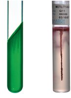
Physical State: Liquid (broth), solid (agar plates), semisolid (motility testing).
Chemical Composition: Defined (precisely known) vs. Complex (not fully defined, e.g., blood, milk).
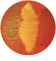
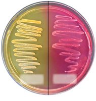
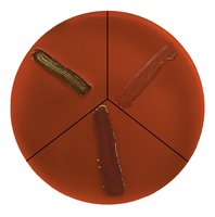
Function: Selective (inhibits some microbes), differential (distinguishes between microbes), or both.
Examples of Differential and Selective Media
Blood Agar: Differentiates based on hemolysis (alpha, beta, gamma).
Mannitol Salt Agar (MSA): Selects for salt-tolerant bacteria; differentiates based on mannitol fermentation.
Eosin Methylene Blue Agar (EMB): Selects against Gram-positive bacteria; differentiates lactose fermenters.
Isolation and Enumeration Techniques
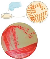
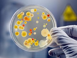
Streak Plate Technique: Used to isolate pure colonies from mixed cultures.
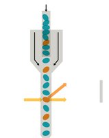
Direct Counting: Manual (microscope, counting chamber), automated (Coulter counter, flow cytometer).
Viable Plate Count: Serial dilution and plating to count colony-forming units (CFU).
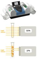
Indirect Methods: Turbidity (spectrophotometry), dry weight, metabolic activity.
Methods for Identifying Microbes
Phenotypic Methods
Microscopic Morphology: Cell shape, arrangement, staining reactions.
Macroscopic Morphology: Colony appearance, growth patterns.
Biochemical Characteristics: Enzyme production, metabolic activities, fermentation tests.
Chemical Analysis: Cell wall composition, lipid profiles.
Immunologic and Genotypic Methods
Immunologic: Detection of specific antigens or antibodies (e.g., ELISA, lateral flow tests).
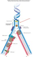
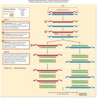
Genotypic: DNA-based identification (e.g., PCR, gene probes, DNA fingerprinting).
Controlling Microbial Growth
Definitions and Principles
Decontamination: Reduces microbial load to safe levels.
Sterilization: Destroys all microbes, including endospores.
Disinfection: Reduces microbial numbers on surfaces or objects.
Microbiostatic: Inhibits microbial growth.
Microbiocidal: Kills microbes.
Disinfectant: Used on inanimate objects.
Antiseptic: Used on living tissue.
Physical Methods of Control
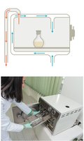
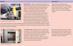
Temperature: Refrigeration slows growth; heat (boiling, autoclaving, dry heat) kills microbes.
Decimal Reduction Time (D value): Time to kill 90% of microbes at a given temperature.
Thermal Death Time: Shortest time to kill all microbes at a set temperature.
Thermal Death Point: Lowest temperature to kill all microbes in 10 minutes.

Radiation and Filtration
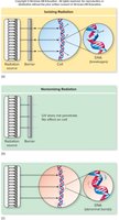
Ionizing Radiation: Gamma rays/X-rays; penetrates packaging, sterilizes heat-sensitive items.
Nonionizing Radiation: UV light; disinfects surfaces, air, and water.
Filtration: HEPA filters for air, membrane filters for liquids; removes microbes without heat.

Chemical Methods: Germicides
Alcohols: Denature proteins, disrupt membranes (e.g., ethanol, isopropanol).
Aldehydes: High/intermediate-level; sterilize equipment (e.g., glutaraldehyde).
Phenols: Disrupt cell walls; used in hygiene products.
Halogens: Oxidize cell components (e.g., chlorine, iodine).
Peroxygens: Strong oxidizers (e.g., hydrogen peroxide).
Ethylene Oxide: Gas sterilant for heat-sensitive materials.
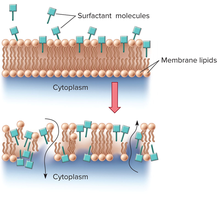
Detergents: Amphipathic molecules; disrupt membranes, remove dirt and microbes.
Special Considerations for Microbial Control
Mycobacteria: Resistant due to waxy cell wall; require strong disinfectants.
Endospores: Highly resistant; best eliminated by autoclaving.
Viruses: Enveloped viruses are sensitive to heat and detergents; naked viruses require stronger agents.
Protozoa: Some stages are resistant; filtration and chemical treatments used.
Prions: Extremely resistant; require combined chemical and physical treatments.
Visual Summary
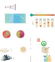
Additional info: These notes provide a comprehensive overview of microbial growth, environmental requirements, laboratory cultivation, enumeration, identification, and control methods, suitable for exam preparation in a college-level microbiology course.
