 Indietro
Gram Stain and Capsule Stain: Microbiology Lab Study Guide
Guida di studio - Note intelligenti
 Appunti personalizzati basati sui tuoi materiali, ampliati con definizioni chiave, esempi e contesto.
Gram Staining and Capsule Staining
Introduction
Gram staining and capsule staining are essential techniques in microbiology for differentiating bacterial cell wall types and visualizing bacterial capsules. These methods provide critical information for identifying bacteria and understanding their structural and functional properties.
Capsule Stain
Definition and Function of Capsules
Capsules are slimy, viscous coverings composed of polysaccharides and polypeptides that surround some bacterial cells. Although not essential for life, capsules confer several advantages:
Resist phagocytosis by host immune cells
Prevent engulfment by protozoans in soil and water
Adhere to surfaces, aiding colonization
Prevent/delay drying (desiccation)
Assist in nutrient gathering
Principle of Capsule Staining
Capsules do not stain well with basic dyes. To visualize capsules, both the bacterial cell and the background are stained, leaving the capsule itself colorless.
Skim milk powder is added to the culture to enhance capsule visibility and adherence to the slide.
Heat fixing is avoided, as it destroys the capsule. Capsules allow cells to stick to the glass without heat.
Capsule Stain Procedure
Add dried skim milk powder to the bacterial culture and mix well.
Place 2-3 loopfuls of the culture on a microscope slide and spread to cover about one inch.
Allow the slide to air dry completely (do not heat fix).
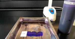
Stain the dried smear with crystal violet for 1 minute.
Wash off excess dye with 20% copper sulfate (CuSO4), which decolorizes the capsule but not the cell.
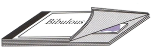
Shake off excess CuSO4 and blot dry.

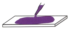
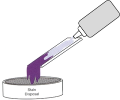
Capsule Stain Results
Bacterial cell and background stain purple; capsule appears colorless.
Both the cell and background are stained, not just one.
Gram Staining
Principle and Purpose
Gram staining is a differential stain used to distinguish between bacteria with gram-positive and gram-negative cell walls. It is the most widely-used staining procedure in bacteriology.
Gram-positive bacteria stain purple.
Gram-negative bacteria stain red/pink.
The terms positive/negative refer to staining results, not electrical charge.
Cell Wall Structure Differences
The staining differences arise from fundamental structural differences in bacterial cell walls:
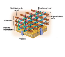
Gram-positive cell wall: 60-90% peptidoglycan, contains teichoic acids.
Gram-negative cell wall: 10-20% peptidoglycan, has an outer membrane with lipopolysaccharides (LPS).
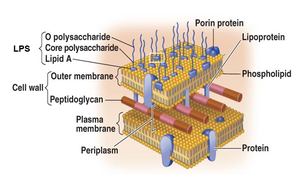
Gram Stain Procedure
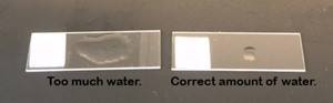
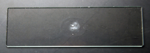
Prepare a bacterial smear on a slide and allow to air dry.
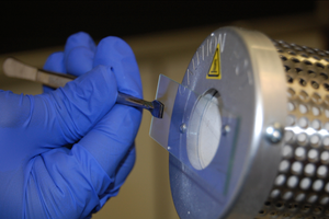
Heat fix the slide by holding it near a microincinerator for 10 seconds.
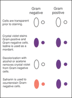
Apply crystal violet (primary stain) for 1 minute, then rinse with water.
Apply Gram’s iodine (mordant) for 1 minute, then rinse with water.
Decolorize with alcohol/acetone until runoff is clear, then rinse with water.
Apply safranin (counterstain) for 1 minute, then rinse with water.
Blot dry with bibulous paper and observe under oil immersion microscopy.
Key Reagents in Gram Staining
Primary stain: Crystal violet
Mordant: Iodine
Decolorizer: Alcohol/acetone
Counterstain: Safranin
Theory Behind Gram Staining
In gram-positive bacteria, crystal violet and iodine form a complex that is trapped by the thick peptidoglycan layer after dehydration by alcohol/acetone.
In gram-negative bacteria, alcohol/acetone dissolves the outer membrane and the thin peptidoglycan layer cannot retain the crystal violet-iodine complex, resulting in decolorization.
Reasons for False Results
Gram-positive cells may stain red/pink if over-heated, over-decolorized, washed excessively, or if the culture is old.
Gram-negative cells may stain purple if the smear is too thick, under-decolorized, or washed insufficiently.
Gram Stain Results and Interpretation
Escherichia coli is gram-negative (red/pink).
Staphylococcus epidermidis is gram-positive (purple).
Mixed stains can show both cell types on the same slide.
Skills Assessment and Study Guide
Essential Skills
Prepare a bacterial smear and perform Gram staining.
Determine whether a culture is Gram positive or Gram negative.
Focus slides under oil immersion and identify cell shape and arrangement.
Identify capsules and differentiate between stained cell, capsule, and background.
Review Questions
What is a capsule? What advantages does it confer?
What do you stain to observe a capsule?
What are the steps of capsule staining?
Why use skim milk powder in capsule staining?
What happens if you heat-fix before capsule staining?
How do cells adhere to the slide without heat fixing?
Is the background, cell, or both stained in capsule staining?
What type of staining method is Gram stain?
What is a differential stain? How is it different from a simple stain?
What cellular structure does Gram staining differentiate?
What color do Gram-positive and Gram-negative bacteria take up?
Describe color changes during Gram staining steps.
Why do Gram-positive and Gram-negative bacteria stain differently?
What are the steps and correct order of Gram staining reagents?
What are the primary stain, mordant, decolorizer, and counterstain?
What results occur if steps are missed or combined incorrectly?
What happens if you decolorize too long?
What are reasons for false results?
Identify Gram-positive and Gram-negative cells in microscopic slides.
Summary Table: Gram Stain Steps and Results
Step | Gram-negative | Gram-positive |
|---|---|---|
Cells prior to staining | Transparent | Transparent |
Crystal violet + iodine | Purple | Purple |
Decolorization | Colorless | Purple |
Safranin | Red/Pink | Purple |
Key Equations and Concepts
Peptidoglycan content: Gram-positive (60-90%), Gram-negative (10-20%)
Gram stain theory: Retention or loss of crystal violet-iodine complex depends on cell wall structure.
Example: Staphylococcus epidermidis (Gram-positive) stains purple; Escherichia coli (Gram-negative) stains red/pink.
