 Indietro
Gram Staining and Broth Culture Techniques in Microbiology
Guida di studio - Note intelligenti
 Appunti personalizzati basati sui tuoi materiali, ampliati con definizioni chiave, esempi e contesto.
Gram Staining and Broth Culture Techniques
Introduction to Gram Staining
The Gram stain is a fundamental differential staining technique in microbiology, developed in 1884 by Hans Christian Gram. It is used to classify bacteria into two major groups—Gram-positive and Gram-negative—based on differences in their cell wall structure. This classification is crucial for bacterial identification and for guiding appropriate antibiotic therapy.
Gram-positive bacteria: Have a thick peptidoglycan layer and retain the primary stain, appearing purple under the microscope.
Gram-negative bacteria: Possess a thin peptidoglycan layer and an outer membrane containing lipopolysaccharide (LPS); they do not retain the primary stain and appear pink after counterstaining.
Clinical importance: The Gram reaction helps determine effective antibiotic treatment, as Gram-negative bacteria are often more resistant due to their outer membrane.
Principle of the Gram Stain
The Gram stain differentiates bacteria based on the physical and chemical properties of their cell walls. The thick peptidoglycan in Gram-positive cells traps the crystal violet-iodine complex, while the outer membrane of Gram-negative cells is disrupted by ethanol, allowing the complex to be washed out and replaced by the pink safranin counterstain.
Key steps: Application of crystal violet, iodine (mordant), ethanol (decolorizer), and safranin (counterstain).
Result: Gram-positive cells remain purple; Gram-negative cells turn pink.
Gram Stain Protocol
The following steps outline the standard Gram staining procedure:
Crystal Violet: Flood the heat-fixed smear with crystal violet for 90 seconds.
Rinse: Briefly rinse with distilled water.
Gram's Iodine: Apply iodine solution for 1 minute (acts as a mordant).
Decolorization: Wash with 95% ethanol until runoff is colorless (about 5 seconds), then rinse immediately with distilled water.
Safranin: Counterstain with safranin for 20 seconds.
Final Rinse and Dry: Rinse gently with distilled water, blot dry with bibulous paper, and air-dry.
Microscopy: Examine under oil immersion at 100× objective.
Expected Results for Common Bacteria
Organism | Gram Reaction | Shape | Color |
|---|---|---|---|
Staphylococcus aureus | Gram-positive | Coccus | Purple |
Bacillus megaterium | Gram-positive | Long bacillus | Purple |
Escherichia coli | Gram-negative | Short bacillus | Pink |
Mycobacterium smegmatis | Gram-positive | Bacillus (tapered ends) | Purple |
Cell Wall Structure Comparison
Feature | Gram-positive | Gram-negative |
|---|---|---|
Peptidoglycan Layer | Thick | Thin |
Outer Membrane | Absent | Present (contains LPS) |
Teichoic Acids | Present | Absent |
Stain Color | Purple | Pink |
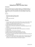
Making Broth Cultures from Slant Cultures
Broth cultures are used to grow bacteria in a liquid medium, which is useful for various assays and for obtaining large numbers of cells. The process involves transferring bacteria from a solid agar slant to a nutrient broth tube using aseptic technique.
Purpose: To prepare bacteria for staining, motility assays, and isolation techniques.
Materials: Slant culture, nutrient broth, inoculating loop, Bunsen burner, and sterile technique supplies.
Procedure for Broth Culture Preparation
Label the broth tube with the organism name and date.
Flame the inoculating loop until red-hot and allow to cool.
Remove the cap from the slant culture and flame the tube's mouth.
Obtain a small amount of growth from the slant with the sterile loop.
Transfer the inoculum into the broth tube, swirl gently.
Flame the mouth of the broth tube and replace the cap.
Incubate the broth tube at the appropriate temperature for 24–48 hours.
Stain and Biohazard Disposal
Crystal violet waste: Dispose in the designated beaker, not the sink.
Other stains (iodine, ethanol, safranin): May be drained into the sink.
Biohazardous materials: Place all contaminated items in biohazard bags; do not dispose of chemicals or biologicals down the sink.
Spill cleanup: Use Bon Ami and water for chemical spills; notify instructor for biological spills.
Microscopy and Drawing Results
After Gram staining, students should observe and draw the four organisms, labeling each with the organism name, Gram reaction, and observed color (purple for Gram-positive, pink for Gram-negative).
Staphylococcus aureus: Gram-positive coccus, purple
Bacillus megaterium: Gram-positive long bacillus, purple
Escherichia coli: Gram-negative short bacillus, pink
Mycobacterium smegmatis: Gram-positive bacillus with tapered ends, purple
Additional info:
Gram-variable reactions may occur, especially with older cultures or certain genera (e.g., Bacillus), but classification should default to Gram-positive if both colors are present.
Proper aseptic technique is essential to avoid contamination during transfers and staining procedures.
