 Indietro
History of Microbiology and Microscopy: Foundations, Key Discoveries, and Tools
Guida di studio - Note intelligenti
 Appunti personalizzati basati sui tuoi materiali, ampliati con definizioni chiave, esempi e contesto.
Introduction to Microbiology
Definition and Scope
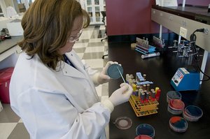
Microbiology is the scientific study of organisms too small to be seen with the naked eye, including bacteria, archaea, fungi, protozoa, helminths, viruses, and prions. This field explores their structure, function, classification, and role in health, disease, and the environment.
Microbes are ubiquitous and play essential roles in ecosystems, industry, and medicine.
Microbiology encompasses both cellular (prokaryotic and eukaryotic) and acellular entities.
Major Groups of Microorganisms
Bacteria and Archaea
Bacteria and archaea are unicellular prokaryotes lacking a nucleus. They are found in diverse environments, including extreme habitats, and reproduce asexually.
Bacteria: Common in soil, water, and living hosts; some are pathogenic.
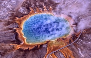
Archaea: Often inhabit extreme environments (e.g., hot springs, salt lakes).
Fungi
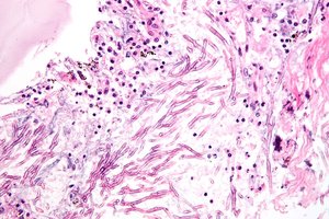
Fungi are eukaryotic organisms that obtain nutrients from other organisms and possess cell walls. They exist as multicellular molds or unicellular yeasts.
Molds: Multicellular, form hyphae, reproduce by spores.
Yeasts: Unicellular, reproduce by budding or spores.
Protozoa (Protists)
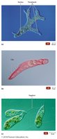
Protozoa are single-celled eukaryotes with animal-like characteristics. They are mostly aquatic, reproduce sexually or asexually, and are often motile.
Examples include Amoeba, Paramecium, and Euglena.
Helminths
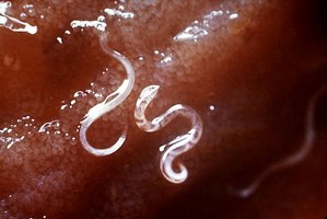
Helminths are parasitic worms, including tapeworms and roundworms. While adult forms are macroscopic, their eggs and larvae are microscopic and studied in microbiology.
Helminths are eukaryotic and multicellular.
Acellular Infectious Agents: Viruses and Prions
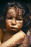
Viruses are acellular, obligate intracellular parasites composed of genetic material and a protein coat. Prions are infectious proteins lacking nucleic acids.
Viruses: Cause diseases such as influenza, HIV/AIDS, and smallpox.
Prions: Cause neurodegenerative diseases like Creutzfeldt-Jakob disease.
Historical Foundations of Microbiology
Spontaneous Generation vs. Biogenesis
Early scientists debated whether life could arise spontaneously (abiogenesis) or only from pre-existing life (biogenesis).
Spontaneous Generation: Proposed by Aristotle; life arises from non-living matter.
Biogenesis: Life arises only from existing life; supported by experiments of Redi, Pasteur, and others.
Redi’s Experiments
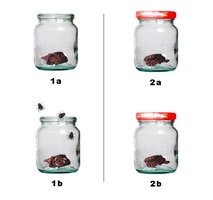
Francesco Redi demonstrated that maggots on decaying meat arose from fly eggs, not spontaneous generation, by using covered and uncovered jars.
Helped disprove abiogenesis for larger organisms.

Antonie van Leeuwenhoek and Robert Hooke
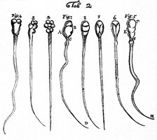
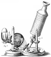
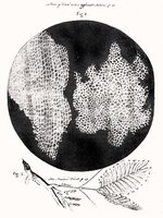
Antonie van Leeuwenhoek improved the microscope and was the first to observe and describe microorganisms, which he called "animalcules." Robert Hooke coined the term "cell" and contributed to cell theory.
Leeuwenhoek: Father of microbiology, bacteriology, and protozoology.
Hooke: First to publish a description of a microorganism (fungus), formulated cell theory.


Louis Pasteur and the Germ Theory of Disease
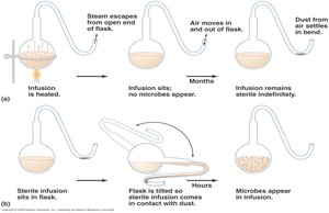
Louis Pasteur disproved spontaneous generation for microbes using swan-neck flask experiments, developed pasteurization, and initiated the germ theory of disease. He also created vaccines for rabies and anthrax.
Demonstrated that microbes cause fermentation and spoilage.
Showed that specific microbes cause specific diseases.
Robert Koch and Koch’s Postulates
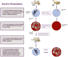
Robert Koch established methods to link specific microbes to specific diseases, known as Koch’s postulates. He isolated the causative agents of anthrax, tuberculosis, and cholera.
Koch’s postulates are foundational for identifying disease-causing organisms.
Joseph Lister and Ignaz Semmelweis: Aseptic Technique
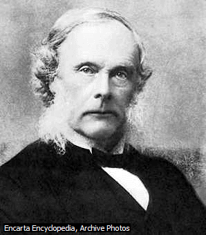
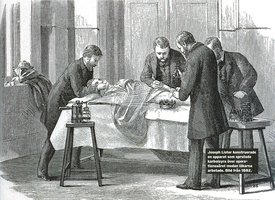
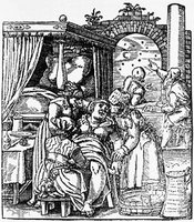
Joseph Lister introduced antiseptic techniques in surgery, including handwashing, sterilization of instruments, and use of disinfectants. Ignaz Semmelweis advocated handwashing to prevent puerperal fever in maternity wards.
These practices greatly reduced infection rates in medical settings.
Microscopy and Microbiological Tools
Principles of Microscopy
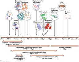
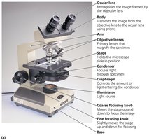
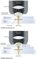
Microscopy is essential for visualizing microorganisms. Key concepts include wavelength, magnification, contrast, and resolution. The compound light microscope is the most common tool in microbiology labs.
Magnification: Enlargement of the specimen image.
Resolution: Ability to distinguish two points as separate.
Contrast: Difference in light intensity between specimen and background.
Types of Microscopes
Several types of microscopes are used in microbiology, each with specific applications and limitations.
Light Microscopes: Bright-field (simple and compound), dark-field, phase-contrast, fluorescence.
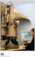
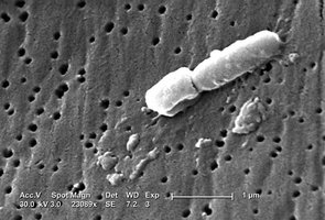
Electron Microscopes: Transmission Electron Microscope (TEM) and Scanning Electron Microscope (SEM) provide much higher magnification and resolution.
Staining Techniques
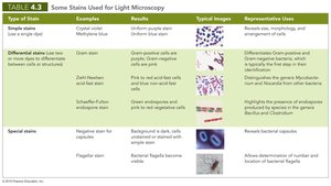
Staining enhances contrast and allows visualization of cellular structures. Stains are classified as simple, differential, or special stains.
Simple stains: Use a single dye to highlight cells.
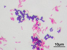
Differential stains: Use multiple dyes to distinguish cell types (e.g., Gram stain, acid-fast stain).
Special stains: Highlight specific structures (e.g., capsule, flagella).
Type of Stain | Examples | Typical Images | Representative Uses |
|---|---|---|---|
Simple stain | Crystal violet, Methylene blue | Uniform purple/blue cells | Reveals cell morphology and arrangement |
Differential stain | Gram stain, Acid-fast stain | Gram-positive (purple), Gram-negative (pink); Acid-fast (pink), Non-acid-fast (blue) | Distinguishes cell wall types; identifies Mycobacterium |
Special stain | Negative (capsule), Flagellar | Background is dark, capsules/flagella visible | Reveals capsules and bacterial flagella |
Bacterial Morphology and Arrangement
Shapes of Bacteria
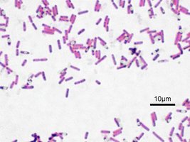
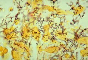
Bacteria exhibit several characteristic shapes, which aid in identification and classification.
Coccus: Spherical
Bacillus: Rod-shaped
Coccobacillus: Short and plump
Vibrio: Gently curved
Spirillum: Spiral-shaped
Spirochete: Corkscrew-shaped
Pleomorphism: Variation in shape and size within a species
Arrangements of Bacteria
Bacterial cells may be arranged in characteristic patterns, depending on the plane of division and whether cells remain attached after division.
Cocci: Single, pairs (diplococci), tetrads, clusters (staphylococci), chains (streptococci), cubical packets (sarcina)
Bacilli: Single, pairs (diplobacilli), chains (streptobacilli), palisades
Summary and Take Home Points
Microbiology studies a diverse group of organisms, both cellular and acellular.
Key historical experiments disproved spontaneous generation and established the germ theory of disease.
Microscopy and staining are essential tools for observing and classifying microbes.
Bacterial morphology and arrangement are important for identification and taxonomy.
Additional info: Students should be able to summarize the contributions of major scientists, design simple experiments to test biogenesis, and explain the principles behind microscopy, staining, and aseptic technique.
