 Indietro
Innate Immunity and Nonspecific Defenses: Mini-Textbook Study Notes
Guida di studio - Note intelligenti
 Appunti personalizzati basati sui tuoi materiali, ampliati con definizioni chiave, esempi e contesto.
Innate Immunity: Overview
Introduction to Host Defenses
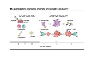
Innate immunity refers to the nonspecific mechanisms that protect the host from infectious diseases. These defenses are present from birth and act rapidly to prevent the entry and spread of pathogens. Unlike adaptive immunity, innate responses do not require prior exposure to the pathogen and are not antigen-specific.
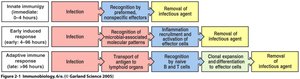
Innate Immunity: Immediate, nonspecific defense mechanisms.
Adaptive Immunity: Specific, delayed response involving lymphocytes and antibodies.
Three Phases of Immune Response: Germline-encoded recognition (innate), early induced response, and antigen-specific adaptive response.
First Line of Defense: Physical and Chemical Barriers
Skin and Mucous Membranes
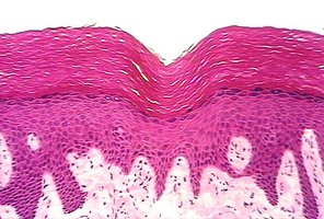
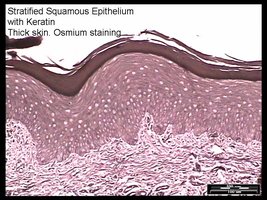
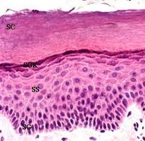
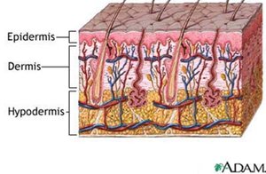
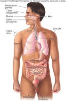
The skin and mucous membranes form the primary physical barriers against pathogens. Their structure and secretions help prevent microbial invasion.
Skin: Composed of tightly packed cells, keratinized layers, and low water content, making it difficult for microbes to penetrate.
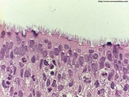
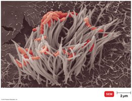
Mucous Membranes: Trap microbes in mucus; ciliated cells help expel pathogens.
Sloughing: Shedding of skin cells removes attached microbes.
Movement of Bodily Fluids
Bodily fluids such as mucus, saliva, and tears help flush pathogens from exposed surfaces. Ciliated cells in the respiratory tract move mucus and trapped microbes out of the body.
Coughing, Sneezing, Vomiting, Diarrhea: Expel pathogens from the body.
Ciliated Cells: Move mucus and pathogens out of the respiratory tract.
Secretions
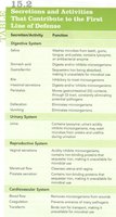
Secretions from various organs contain antimicrobial substances that inhibit pathogen growth.
Lysozyme: Enzyme in sweat, tears, and saliva that destroys bacterial cell walls.
Acidic Environment: Stomach acid (pH 1.2–3.0), skin (pH 3–5), and vaginal secretions create hostile conditions for microbes.
Fatty Acids: Sebum and oil secreted by skin are fungistatic and bacteriostatic.
Normal Flora
Normal microbiota outcompete pathogens for resources and produce substances that inhibit pathogen survival. This is known as microbial antagonism.
Microbial Antagonism: Normal flora produce bacteriocins and alter environmental conditions.
Population: Over 100 trillion bacteria inhabit the human body, compared to 10 trillion human cells.
Antimicrobial Peptides and Chemicals
Antimicrobial peptides are triggered by microbial sugars and proteins and can have some specificity. Chemical factors include fungistatic fatty acids, lysozyme, and acidic pH in various secretions.
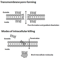
Peptides: Defensins, cathelicidins, and others disrupt microbial membranes.
Chemical Factors: Low pH, lysozyme, fatty acids.
Second Line of Defense: Internal Nonspecific Mechanisms
Complement System
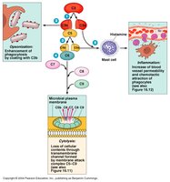
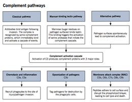
The complement system consists of at least 26 blood proteins that work together to destroy bacteria and viruses. Activation occurs via three pathways: classical, lectin, and alternative.
Activation: Antigen-antibody reaction or direct pathogen recognition.
Functions (COLA): Chemotaxis, Opsonization, Lysis, Activation of inflammation.
Membrane Attack Complex (MAC): Forms pores in microbial membranes, causing lysis.
Phagocytosis
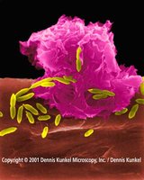
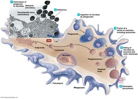
Phagocytosis is the process by which phagocytes ingest and destroy pathogens. Major phagocytes include macrophages, neutrophils, and eosinophils.
Stages (C.A.I.D.): Chemotaxis, Adherence, Ingestion, Digestion.
Opsonization: Complement proteins or antibodies enhance adherence.
Microbial Evasion: Some bacteria, like Mycobacterium spp., resist killing by phagocytes.
Cells of the Second Line of Defense
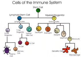
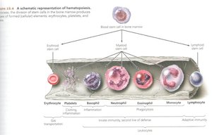
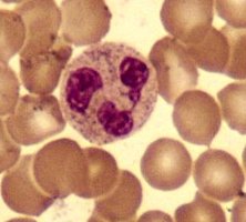
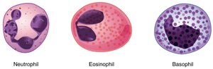
Granulocytes and agranulocytes are key players in innate immunity. Granulocytes include neutrophils, eosinophils, and basophils. Agranulocytes include monocytes (which mature into macrophages and dendritic cells) and lymphocytes.
Neutrophils: Phagocytosis, production of reactive oxygen species.
Eosinophils: Attack large eukaryotic pathogens, involved in inflammation and allergies.
Basophils: Similar to eosinophils, involved in allergic responses.
Monocytes: Mature into macrophages and dendritic cells.
Lymphocytes: Part of the adaptive immune response.
Natural Killer (NK) Cells
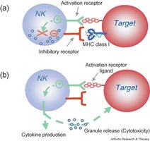
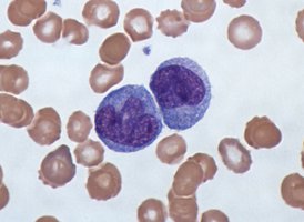
Natural killer cells are lymphocytes that can destroy infected or tumor cells without prior antigen exposure. They recognize abnormal cells and induce apoptosis.
Targeting: NK cells recognize cells lacking normal MHC I molecules.
Action: Release cytotoxic granules and cytokines.
Interferons
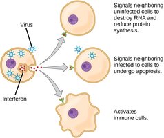
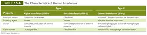
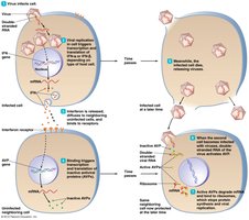
Interferons are signaling proteins released by cells in response to viral infection. They activate antiviral defenses in neighboring cells and modulate immune responses.
Types: IFN-α, IFN-β (Type I); IFN-γ (Type II).
Actions: Induce antiviral proteins, activate phagocytes, regulate immune communication.
Fever
Fever is a systemic response to infection, mediated by interleukins and prostaglandins. It enhances immune function and inhibits pathogen growth.
Mechanism: Interleukins stimulate hypothalamus to release prostaglandins, raising body temperature.
Benefits: Inhibits microbial growth, increases immune cell activity, speeds up chemical reactions, reduces endotoxin effects.
Inflammation
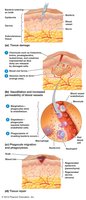
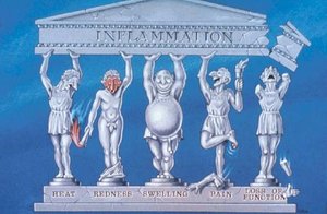
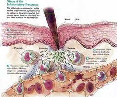
Inflammation is the body's response to injury or infection, characterized by redness, heat, swelling, and pain. It involves increased blood flow, vascular permeability, and recruitment of immune cells.
Acute vs. Chronic: Acute inflammation is short-term; chronic is prolonged and may cause tissue damage.
Symptoms: Redness (rubor), heat (calor), swelling (tumor), pain (dolor).
Causes: Blood flow, chemical mediators, fluid leakage, nerve injury.
Summary Table: Nonspecific Components of Innate Immunity
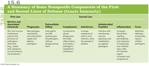
The following table summarizes the main nonspecific components of the first and second lines of defense:
First Line | Second Line |
|---|---|
Barriers & Associated Chemicals | Phagocytes, Extracellular Killing, Complement, Interferons, Antimicrobial Peptides, Inflammation, Fever |
Physical barriers, secretions, normal flora | Macrophages, NK cells, complement proteins, interferons, peptides, inflammatory mediators, elevated temperature |
Take Home Points
Understand the five factors in the first line of defense (physical barriers).
Know the six nonspecific tools of the second line of defense.
Learn the steps of phagocytosis.
Comprehend the functions of NK cells and interferons.
Recognize the four functions of complement proteins.
Identify the four symptoms of inflammation and their causes.
