 Indietro
Innate Immunity: Nonspecific Defenses of the Host – Study Notes
Guida di studio - Note intelligenti
 Appunti personalizzati basati sui tuoi materiali, ampliati con definizioni chiave, esempi e contesto.
Innate Immunity: Nonspecific Defenses of the Host
The Concept of Immunity
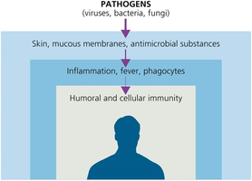
Immunity refers to the ability of an organism to resist infection or disease. The immune system is divided into two main types: innate immunity and adaptive immunity. Innate immunity is present at birth and provides rapid, nonspecific defense against pathogens, while adaptive immunity is specific and develops more slowly, featuring memory for previously encountered pathogens.
Immunity: Ability to ward off disease.
Susceptibility: Lack of resistance to a disease.
Innate immunity: Defenses against any pathogen; rapid, present at birth.
Adaptive immunity: Immunity or resistance to a specific pathogen; slower to respond, has memory component.
Toll-like receptors (TLRs) on host cells recognize pathogen-associated molecular patterns (PAMPs) such as LPS, flagellin, peptidoglycan, and viral nucleic acids. Binding of TLRs to PAMPs triggers the release of cytokines, which regulate immune responses and recruit other immune cells.
Physical Factors of Innate Immunity
Physical barriers are the first line of defense against pathogens. These include the skin, mucous membranes, and various mechanical processes that remove microbes from the body.
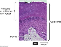
Skin: Composed of the dermis (connective tissue) and epidermis (tightly packed epithelial cells with keratin). Shedding and dryness inhibit microbial growth.
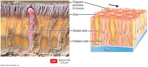
Mucous membranes: Line the gastrointestinal, respiratory, and genitourinary tracts. Mucus traps microbes and prevents drying.
Lacrimal apparatus: Drains tears and washes the eye.
Ciliary escalator: Transports microbes trapped in mucus away from the lungs.
Epiglottis: Prevents microorganisms from entering the lower respiratory tract.
Earwax, urine, vaginal secretions, peristalsis, defecation, vomiting, diarrhea: Mechanisms to expel microbes.
Chemical Factors of Innate Immunity
Chemical barriers complement physical defenses by creating hostile environments for pathogens.
Sebum: Forms a protective film and lowers skin pH (3–5).
Lysozyme: Enzyme in perspiration, tears, saliva, and urine that destroys bacterial cell walls.
Gastric juice: Low pH (1.2–3.0) destroys most bacteria and toxins.
Vaginal secretions: Low pH (3–5) inhibits microbes.
Normal Microbiota and Innate Immunity
Normal microbiota play a crucial role in protecting the host by competing with pathogens and influencing immune development.
Microbial antagonism: Normal microbiota compete for space and nutrients, produce substances harmful to pathogens, and alter conditions affecting pathogen survival.
Commensalism: One organism benefits, the host is unharmed.
Opportunistic pathogens: Some normal microbiota can cause disease under certain conditions (e.g., E. coli, S. aureus).
Probiotics: Live microbial cultures administered for beneficial effects.
Prebiotics: Nutrients that promote growth of beneficial bacteria.
Formed Elements in Blood
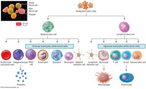
Blood contains various formed elements, including white blood cells (leukocytes), which are essential for immune defense. Leukocytes are classified as granulocytes or agranulocytes based on the presence of cytoplasmic granules.
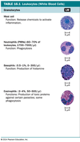
Granulocytes: Neutrophils (phagocytic, early infection), basophils (release histamine, allergic responses), eosinophils (phagocytic, toxic against parasites).
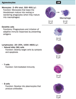
Agranulocytes: Monocytes (mature into macrophages), dendritic cells (phagocytic, initiate immune responses), lymphocytes (T cells, B cells, NK cells).
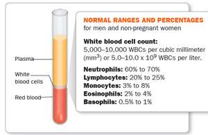
Leukocyte Type | Function | Normal Range (%) |
|---|---|---|
Neutrophils | Phagocytosis; early infection response | 60–70% |
Lymphocytes | Adaptive immunity (T, B, NK cells) | 20–25% |
Monocytes | Mature into macrophages; phagocytosis | 3–8% |
Eosinophils | Toxic against parasites; some phagocytosis | 2–4% |
Basophils | Release histamine; allergic responses | 0.5–1% |
The Lymphoid System
The lymphoid system consists of lymph plasma, lymphatic vessels, and organs containing lymphoid tissue. It is essential for immune surveillance and response.
Lymphoid tissue: Contains lymphocytes and phagocytic cells.
Lymph: Carries microbes to lymph nodes for destruction by B and T lymphocytes, macrophages, and dendritic cells.
Phagocytes and Phagocytosis
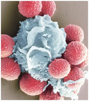
Phagocytosis is the process by which cells ingest and destroy microorganisms or other particles. Professional phagocytes include neutrophils, eosinophils, and macrophages.
Neutrophils: First responders in infection.
Macrophages: Clean-up crew, dominate later in infection.
Fixed macrophages: Reside in tissues and organs.
Free macrophages: Roam tissues and gather at infection sites.
Mechanism of Phagocytosis
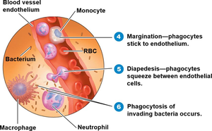
Chemotaxis: Chemical signals attract phagocytes to microorganisms.
Adherence: Phagocyte attaches to the microorganism; opsonization enhances adherence.
Ingestion: Pseudopods engulf the microorganism, forming a phagosome.
Digestion: Lysosomes fuse with phagosome, forming a phagolysosome; enzymes and toxic oxygen products digest the microorganism.
Exocytosis: Indigestible material is expelled from the cell.
Inflammation
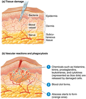
Inflammation is a local defensive response triggered by tissue damage. It serves to destroy or limit the effects of injurious agents and repair damaged tissue.
Signs and symptoms (PRISH): Pain, redness, immobility, swelling (edema), heat.
Acute inflammation: Rapid onset, short duration.
Chronic inflammation: Slow onset, long duration, may be severe.
Stages of Inflammation
Vasodilation and increased vascular permeability: Blood vessels dilate and become more permeable, allowing defensive substances to reach the injured area.
Phagocyte migration and phagocytosis: Phagocytes move to the site of injury and ingest microbes.
Tissue repair: Damaged tissue is repaired and replaced.
Fever
Fever is an abnormally high body temperature, often caused by infection. It is a defense mechanism that enhances immune function and inhibits pathogen growth.
Causes: Bacterial infection, toxins, viral infections.
Mechanism: Cytokines stimulate the hypothalamus to raise body temperature.
Benefits: Enhances phagocyte and T cell activity, intensifies antimicrobial substances, slows pathogen growth, speeds repair.
Complications: Tachycardia, acidosis, dehydration, seizures, coma.
The Complement System
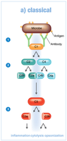
The complement system consists of over 30 serum proteins that enhance the immune response by destroying microbes through cytolysis, opsonization, and inflammation. Complement activation occurs in a cascade and can be triggered by both innate and adaptive immunity.
Classical pathway: Antibodies bind to antigens, activating C1, which splits and activates C2 and C4. C2a and C4b combine to activate C3. C3a promotes inflammation; C3b promotes cytolysis and opsonization.
Outcomes: Cytolysis (membrane attack complex), opsonization (enhanced phagocytosis), inflammation (histamine release).
Outcome | Description |
|---|---|
Cytolysis | Membrane attack complex (MAC) forms, lysing microbes |
Opsonization | Phagocyte attachment to microbe is promoted |
Inflammation | Complement proteins trigger histamine release |
Interferons
Interferons are cytokines produced by host cells in response to viral infection. They induce nearby cells to produce antiviral proteins, inhibit viral replication, and activate immune cells.
Types: Alpha, beta, and gamma interferons.
Functions: Induce antiviral proteins, activate neutrophils and macrophages.
Iron-Binding Proteins
Iron-binding proteins restrict the availability of iron to pathogens, inhibiting their growth.
Transferrin: Blood and tissue fluids.
Lactoferrin: Milk, saliva, mucus.
Ferritin: Liver, spleen, bone marrow.
Hemoglobin: Red blood cells.
Siderophores: Bacterial proteins that compete for iron.
Antimicrobial Peptides
Antimicrobial peptides are short proteins produced in response to microbial molecules. They have broad-spectrum activity and inhibit cell wall synthesis, form pores in membranes, and destroy nucleic acids.
Examples: Dermcidin (skin), defensins, cathelicidins (neutrophils, macrophages), thrombocidin (platelets).
Functions: Inhibit cell wall synthesis, cause lysis, destroy DNA and RNA.
