 Indietro
Innate Immunity: The Body’s First and Second Lines of Defense
Guida di studio - Note intelligenti
 Appunti personalizzati basati sui tuoi materiali, ampliati con definizioni chiave, esempi e contesto.
Innate Immunity: The Body’s First and Second Lines of Defense
The Body’s First Line of Defense
The first line of defense consists of physical and chemical barriers that prevent pathogens from entering the body. These include the skin, mucous membranes, and associated structures and chemicals.
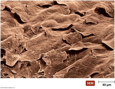
Skin
Structure: The skin is composed of two layers: the epidermis (outer layer) and the dermis (inner layer).
Physical Barrier: The tightly packed cells of the epidermis prevent pathogen entry. Continual shedding removes attached microorganisms.
Chemical Defenses:
Perspiration: Contains salt (inhibits pathogen growth), antimicrobial peptides, and lysozyme (destroys bacterial cell walls).
Sebum: Secreted by sebaceous glands, keeps skin pliable and lowers pH, inhibiting many bacteria.
Example: Lysozyme in sweat and tears breaks down peptidoglycan in bacterial cell walls.
Mucous Membranes
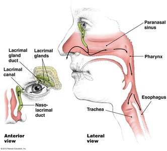
Structure: Line all body cavities open to the environment; consist of epithelium and deeper connective tissue.
Physical Barrier: Tightly packed, living epithelial cells prevent pathogen entry. Continual shedding removes microbes.
Chemical Defenses: Mucus traps microbes; contains antimicrobial chemicals.
Example: Mucous membranes in the respiratory, digestive, urinary, and reproductive systems.

Lacrimal Apparatus
Function: Produces and drains tears, washing the surface of the eye.
Chemical Defense: Tears contain lysozyme, which breaks down peptidoglycan in Gram-positive bacteria.
Note: Most eye infections are caused by Gram-negative bacteria.
Normal Microbiota and Innate Immunity
Microbial Antagonism: Normal microbiota compete with potential pathogens for nutrients and space.
Benefits:
Consume nutrients, making it difficult for pathogens to compete.
Create an unfavorable environment for other microorganisms.
Stimulate the body’s second line of defense.
Promote health by providing vitamins to the host.
Antimicrobial Peptides
Location: Found in skin, mucous membranes, and neutrophils.
Function: Act against a variety of microbes by disrupting membranes or interfering with microbial processes.
The Body’s Second Line of Defense
The second line of defense is activated when pathogens penetrate the skin or mucous membranes. It includes cellular and chemical components, many of which are found in the blood.
Defense Components of Blood
Plasma: Mostly water, contains electrolytes, dissolved gases, nutrients, and proteins (including iron-binding compounds, complement proteins, and antibodies).
Serum: Plasma without clotting factors (fibrinogen).
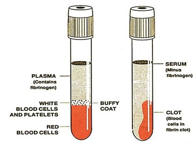
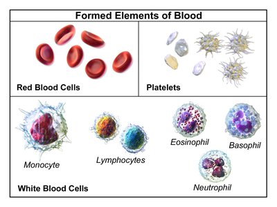
Formed Elements: Cells and cell fragments in plasma. Three main types:
Erythrocytes: Red blood cells, carry oxygen and carbon dioxide.
Platelets: Involved in blood clotting.
Leukocytes: White blood cells, defend against invaders. Divided into granulocytes and agranulocytes.
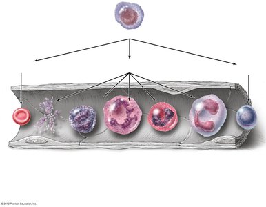
Hematopoiesis
Definition: The process of production, multiplication, and specialization of blood cells in the bone marrow.
Stem Cells: Blood stem cells differentiate into erythroid, myeloid, and lymphoid lineages.
Granulocytes
Definition: White blood cells with granules that stain different colors.
Types:
Basophils: Stain blue with basic dye.
Eosinophils: Stain red/orange with acidic dye.
Neutrophils: Stain lilac with mixed dyes.
Functions: Neutrophils and eosinophils phagocytize pathogens and can kill bacteria by non-phagocytic means. Both are capable of diapedesis (movement through tissues).
Agranulocytes
Definition: White blood cells with uniform cytoplasm.
Types:
Lymphocytes: Most involved in adaptive immunity.
Monocytes: Mature into macrophages in tissues; phagocytic cells.
Lab Analysis of Leukocytes
Differential White Blood Cell Count: Used to signal signs of disease.
Increased eosinophils indicate allergies or parasitic worm infection.
Bacterial diseases often show increased leukocytes and neutrophils.
Viral infections show increased lymphocytes.
Phagocytosis
Definition: The process by which phagocytes ingest and destroy pathogens.
Stages:
Chemotaxis
Adherence
Ingestion
Killing
Elimination
Example: Neutrophils and macrophages are primary phagocytes.

Nonphagocytic Killing
Eosinophils: Attack parasitic helminths by secreting toxins; elevated eosinophils indicate helminth infestation.
Neutrophils: Produce chemicals and extracellular fibers (NETs) to kill bacteria.
Natural Killer (NK) Cells: Secrete toxins onto virally infected cells and tumors; recognize normal cells by membrane proteins.
Chemical Defenses Against Pathogens
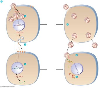
Interferons
Definition: Proteins released by virally infected cells to inhibit viral spread.
Types:
Type I (alpha and beta)
Type II (gamma)
Mechanism: Interferons induce production of antiviral proteins (AVPs) in neighboring cells, which block viral replication.
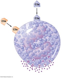
Complement System
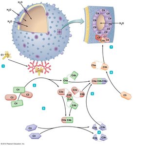
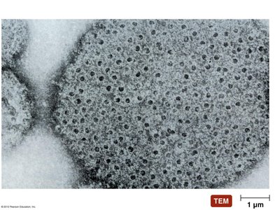
Definition: A set of serum proteins that, when activated, lead to lysis of foreign cells.
Activation Pathways:
Classical pathway
Alternative pathway
Lectin pathway
Functions:
Trigger inflammation
Attract phagocytes
Kill microbes via membrane attack complex (MAC)
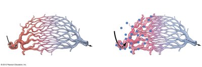
Inflammation
Definition: Nonspecific response to tissue damage.
Characteristics: Redness, heat, swelling, pain.
Types:
Acute: Develops quickly, short-lived, beneficial.
Chronic: Long-lasting, may be harmful.
Mechanisms: Dilation and increased permeability of blood vessels, migration of phagocytes, tissue repair.
Fever
Definition: Body temperature above 37°C, triggered by pyrogens.
Mechanism: Pyrogens stimulate the hypothalamus to increase core temperature. Types of pyrogens include bacterial toxins, cytoplasmic contents released by lysis, and antibody-antigen complexes.
Effects: Increased metabolic rate, shivering, inhibition of sweating, vasoconstriction.
Summary Table: Defense Components of Blood
Component | Function |
|---|---|
Plasma | Transport, contains proteins and nutrients |
Serum | Plasma minus clotting factors |
Erythrocytes | Gas transport |
Platelets | Blood clotting |
Leukocytes | Defense against invaders |
Granulocytes | Phagocytosis, inflammation |
Agranulocytes | Adaptive immunity, phagocytosis |
Summary Table: Stages of Phagocytosis
Stage | Description |
|---|---|
Chemotaxis | Movement toward chemical signals |
Adherence | Attachment to microbe |
Ingestion | Engulfment of microbe |
Killing | Destruction by enzymes and chemicals |
Elimination | Exocytosis of debris |
Key Equations
Fever Mechanism:
Complement Activation (Classical Pathway):
Additional info: Academic context was added to clarify the mechanisms and functions of innate immunity components, as well as to provide self-contained explanations for exam preparation.
