 Indietro
Introduction to Eukaryotic Cells: Structure, Function, and Classification
Guida di studio - Note intelligenti
 Appunti personalizzati basati sui tuoi materiali, ampliati con definizioni chiave, esempi e contesto.
Introduction to Eukaryotic Cells
Overview of Eukaryotes
Eukaryotic cells are fundamental units of life in organisms such as plants, animals, fungi, and protists. They are distinguished from prokaryotic cells by their structural complexity and the presence of membrane-bound organelles.
Endosymbiotic theory: Explains the origin of eukaryotic cells through cell-merging events between ancient eukaryotes and prokaryotes.
Key learning objectives: Describe endosymbiotic theory, compare eukaryotic and prokaryotic cells, contrast mitosis and meiosis, and explain eukaryotic cell transport mechanisms.
The Endosymbiotic Theory
How Eukaryotes Evolved
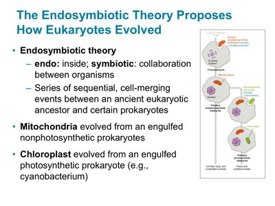
The endosymbiotic theory proposes that eukaryotic cells evolved from a series of symbiotic relationships between ancestral eukaryotes and prokaryotes. Mitochondria and chloroplasts are believed to have originated from engulfed prokaryotes.
Mitochondria: Evolved from engulfed nonphotosynthetic prokaryotes.
Chloroplasts: Evolved from engulfed photosynthetic prokaryotes (e.g., cyanobacteria).
Eukaryotic vs. Prokaryotic Cells
Cell Structures and Processes
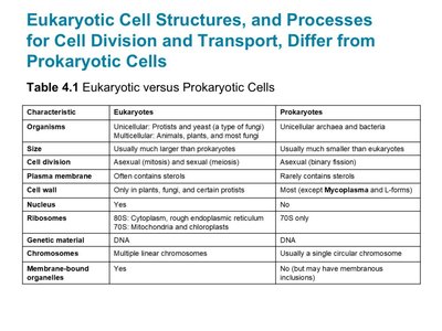
Eukaryotic cells differ from prokaryotic cells in size, complexity, genetic material, and the presence of membrane-bound organelles.
Characteristic | Eukaryotes | Prokaryotes |
|---|---|---|
Organisms | Unicellular (protists, yeast), multicellular (animals, plants, fungi) | Unicellular archaea and bacteria |
Size | Usually larger | Usually smaller |
Cell division | Asexual (mitosis), sexual (meiosis) | Asexual (binary fission) |
Plasma membrane | Often contains sterols | Rarely contains sterols |
Cell wall | Only in plants, fungi, certain protists | Most (except Mycoplasma and L-forms) |
Nucleus | Yes | No |
Ribosomes | 80S (cytoplasm, rough ER), 70S (mitochondria, chloroplasts) | 70S only |
Genetic material | DNA | DNA |
Chromosomes | Multiple linear chromosomes | Single circular chromosome |
Membrane-bound organelles | Yes | No |
Membrane-Bound Organelles
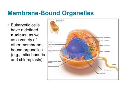
Eukaryotic cells contain a defined nucleus and various membrane-bound organelles, such as mitochondria and chloroplasts, which perform specialized functions.
Cell Division: Mitosis and Meiosis
Mitosis
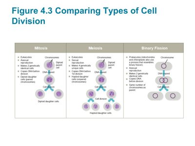
Mitosis is a process of cell division that produces two genetically identical offspring from one parent cell, maintaining the same chromosome number.
Occurs in somatic cells
Ensures genetic stability
Meiosis
Meiosis is involved in sexual reproduction and consists of two cell division stages, resulting in four haploid gametes. Crossing over during meiosis allows for genetic recombination.
Produces gametes (sperm and egg cells)
Gametes are haploid
Crossing over increases genetic diversity
Eukaryotic Cell Transport Mechanisms
Endocytosis and Exocytosis
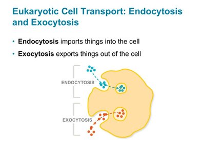
Eukaryotic cells use endocytosis to import substances and exocytosis to export substances. These processes are essential for nutrient uptake, waste removal, and cellular communication.
Endocytosis: Imports materials into the cell.
Exocytosis: Exports materials out of the cell.
Phagocytosis
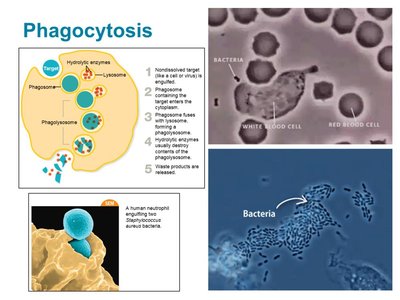
Phagocytosis is a specialized form of endocytosis where cells engulf large particles, such as microbes or cell debris. The process involves the formation of a phagosome, fusion with a lysosome, and digestion of the target.
Performed by specialized immune cells (e.g., macrophages)
Critical for defense against pathogens
Receptor-Mediated Endocytosis and Exocytosis
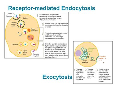
Receptor-mediated endocytosis involves the selective uptake of substances via cell-surface receptors and clathrin-coated vesicles. Exocytosis is the process by which vesicles deliver their contents to the cell surface for secretion or membrane renewal.
Ligands bind to receptors, triggering vesicle formation
Clathrin-coated pits facilitate vesicle budding
Exocytosis replenishes membrane and releases cell products
Classification of Eukaryotes
Four Kingdoms of Eukaryotes
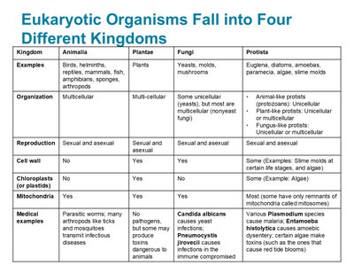
Eukaryotic organisms are classified into four kingdoms: Animalia, Plantae, Fungi, and Protista. Each kingdom has distinct characteristics, modes of reproduction, and medical relevance.
Kingdom | Examples | Organization | Reproduction | Cell wall | Chloroplasts | Mitochondria | Medical examples |
|---|---|---|---|---|---|---|---|
Animalia | Birds, helminths, reptiles, mammals, fish, arthropods | Multicellular | Sexual and asexual | No | No | Yes | Parasitic worms, arthropods, many diseases |
Plantae | Plants | Multicellular | Sexual and asexual | Yes | Yes | Yes | Few pathogens, some infectious agents |
Fungi | Yeasts, molds, mushrooms | Unicellular or multicellular | Sexual and asexual | Yes | No | Yes | Candida, Pneumocystis, dermatophytes |
Protista | Euglena, diatoms, amoebas, paramecia, algae, slime molds | Unicellular, multicellular, multinucleated | Sexual and asexual | Some | Some | Most | Plasmodium, Giardia, Toxoplasma |
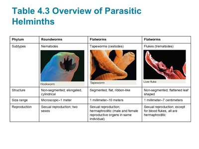
Parasitic Helminths
Helminths are parasitic worms classified as roundworms (nematodes) and flatworms (cestodes and trematodes). They have complex life cycles and are significant causes of infectious diseases worldwide.
Phylum | Subtypes | Structure | Size range | Reproduction |
|---|---|---|---|---|
Roundworms | Nematodes | Non-segmented, elongated, cylindrical | Microscopic–1 meter | Sexual, two sexes |
Flatworms | Tapeworms (cestodes) | Segmented, flat, ribbon-like | 1 mm–10 meters | Sexual, hermaphroditic |
Flatworms | Flukes (trematodes) | Non-segmented, flattened leaf-shaped | 1 mm–7 cm | Sexual, hermaphroditic |
Plants and Fungi
Plants
Plants are multicellular organisms that carry out photosynthesis using chloroplasts. They produce organic carbon from light energy and can serve as vehicles for infectious pathogens.
Fungi
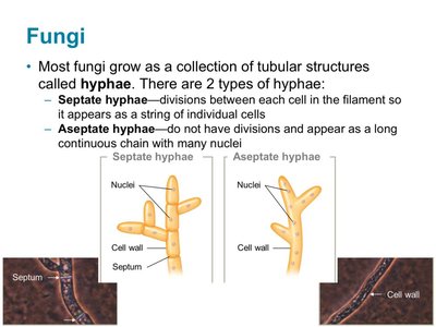
Fungi are a diverse group, most of which are multicellular except for yeasts. They absorb nutrients from their environment and include both pathogens and saprobes. Fungi grow as hyphae, which can be septate or aseptate.
Septate hyphae: Have divisions between cells.
Aseptate hyphae: Lack divisions, forming continuous chains.
Fungal Spores
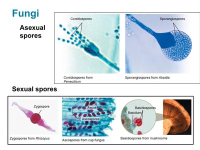
Fungi produce spores for reproduction and classification. Asexual spores arise from mitosis, while sexual spores arise from meiosis.
Asexual spores: Conidiospores, sporangiospores
Sexual spores: Zygospores, ascospores, basidiospores
Fungal Diseases
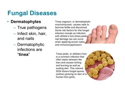
Dermatophytes are true fungal pathogens that infect skin, hair, and nails, causing diseases known as tinea (e.g., athlete's foot, nail infections).
Protists
Overview and Classification
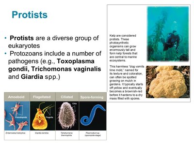
Protists are a diverse group of eukaryotes, including protozoans and algae. Protozoans are classified by their means of motility: amoeboid, flagellated, ciliated, and spore-forming (apicomplexa).
Amoeboid: Move using pseudopods
Flagellated: Move using flagella
Ciliated: Move using cilia
Spore-forming: Move by gliding, complex life cycles
Spore-Forming Protozoans (Apicomplexa)
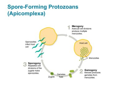
Apicomplexans undergo three life stages: merogony (asexual reproduction), gamogony (sexual reproduction), and sporogony (spore formation). Many are important human pathogens, such as Plasmodium (malaria).
Extracellular Structure
Plasma Membrane and Cell Wall
Eukaryotic plasma membranes contain sterols and are structurally distinct from prokaryotic membranes. Cell walls are present in plants, fungi, and some protists, but absent in animals.
Glycocalyx
The eukaryotic glycocalyx is a carbohydrate-rich layer external to the plasma membrane, serving roles in protection, adhesion, and cellular communication.
Flagella and Cilia
Eukaryotic flagella and cilia are composed of microtubules arranged in a nine-plus-two pattern. They differ from prokaryotic flagella in structure and movement.
Flagella: Long, whip-like structures for movement
Cilia: Short, hair-like structures for movement and sensory functions
Intracellular Structures
Ribosomes
Eukaryotic ribosomes are 80S in the cytoplasm and rough ER, while mitochondria and chloroplasts contain 70S ribosomes similar to prokaryotes. Ribosomes are essential for protein synthesis.
Cytoskeleton
The cytoskeleton is composed of microtubules, intermediate filaments, and microfilaments, providing structural support and facilitating intracellular transport.
Microtubules: Hollow tubes, form spindle during cell division
Intermediate filaments: Provide mechanical strength
Microfilaments: Involved in cell movement and shape
Nucleus
The nucleus is a double-membrane organelle housing the cell's genome. It contains chromatin, nucleoplasm, and the nucleolus, which is the site of ribosome assembly.
Endoplasmic Reticulum and Golgi Apparatus
The endoplasmic reticulum (ER) is involved in protein and lipid production. The rough ER is studded with ribosomes, while the smooth ER is involved in lipid synthesis and detoxification. The Golgi apparatus modifies, sorts, and distributes cellular products.
Mitochondria and Chloroplasts
Mitochondria are the powerhouses of the cell, generating ATP through cellular respiration. Chloroplasts are found in plants and algae, conducting photosynthesis.
