 Indietro
Introduction to Microbiology: Foundations, Methods, and Applications
Guida di studio - Note intelligenti
 Appunti personalizzati basati sui tuoi materiali, ampliati con definizioni chiave, esempi e contesto.
Introduction to Microbiology
Overview of Microbiology
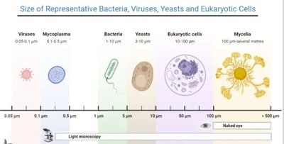
Microbiology is the study of organisms too small to be seen with the unaided eye, known as microorganisms or microbes. This field explores the diversity, structure, growth, genetics, and roles of microbes in health, disease, and the environment. Microbiology is foundational for understanding infectious diseases, biotechnology, and the human microbiome.
Microorganisms include bacteria, archaea, protists, fungi, helminths, viruses, and prions.
Microbes are ubiquitous—found in every environment on Earth.
Most microbes are beneficial or neutral; only a small fraction are pathogenic (disease-causing).
Characteristics of Life
All living things share certain characteristics, though some infectious agents like viruses and prions are exceptions.
Organization: Composed of cells with specialized functions.
Homeostasis: Maintain a stable internal environment.
Growth: Increase in size or number.
Reproduction: Ability to produce offspring.
Metabolism: Chemical reactions for energy and synthesis.
Response to stimuli: React to environmental changes.
Adaptation and evolution: Genetic changes that enhance survival.
History and Themes of Microbiology
Development of Microbiology
The field of microbiology emerged with the invention of the microscope and the observation of microorganisms. Early scientists challenged prevailing theories about disease and life’s origins.
Robert Hooke and Antonie van Leeuwenhoek were pioneers in microscopy, enabling the first observations of microbes.
Spontaneous generation was the belief that life could arise from nonliving matter, later disproven by experiments.
Biogenesis is the principle that life arises from pre-existing life.
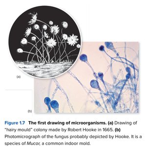
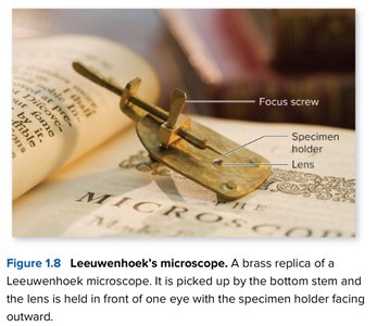
Spontaneous Generation vs. Biogenesis
Experiments by Francesco Redi and Louis Pasteur provided evidence against spontaneous generation, supporting biogenesis.
Redi’s experiment: Showed that maggots on meat came from flies, not spontaneous generation.
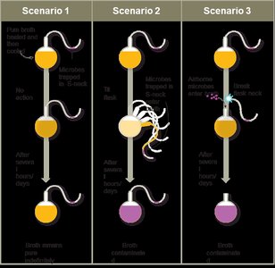
Pasteur’s swan-neck flask experiment: Demonstrated that microbes come from the environment, not from nonliving matter.
The Germ Theory of Disease
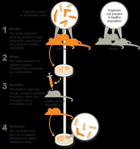
The germ theory established that specific microorganisms cause specific diseases. Robert Koch developed a set of criteria (Koch’s postulates) to link microbes to diseases.
Koch’s postulates: Steps to prove a microbe causes a disease.
Anthrax was shown to be caused by Bacillus anthracis.
Classification and Taxonomy of Microorganisms
Major Groups of Microbes
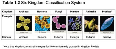
Microbes are classified based on cell type, structure, and genetic relationships.
Microbe | Cell Type | Notes |
|---|---|---|
Bacteria | Prokaryotic | Unicellular; pathogenic and nonpathogenic |
Archaea | Prokaryotic | Unicellular; nonpathogenic; extremophiles |
Protists | Eukaryotic | Unicellular/multicellular; pathogenic and nonpathogenic |
Fungi | Eukaryotic | Unicellular/multicellular; includes yeasts and molds |
Helminths | Eukaryotic | Multicellular; parasitic worms |
Viruses | Not cells | Nonliving; DNA or RNA genome |
Prions | Not cells | Infectious proteins |
Taxonomic Hierarchy and Nomenclature
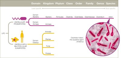
Organisms are classified using a hierarchical system: Domain, Kingdom, Phylum, Class, Order, Family, Genus, Species. Scientific names use binomial nomenclature (Genus species, italicized).
Domains: Bacteria, Archaea, Eukarya
Species: For eukaryotes, organisms that can reproduce; for prokaryotes, those with similar morphology and genetic identity.
Strains: Genetic variants within a species.
Microbes and Human Health
Symbiosis and the Human Microbiome
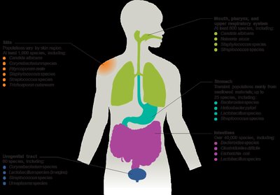
Humans coexist with a vast array of microbes, many of which are essential for health. The human microbiome includes bacteria, archaea, and eukaryotic microbes living on and in the body.
Mutualism: Both host and microbe benefit.
Commensalism: Microbe benefits, host is unaffected.
Parasitism: Microbe benefits at the host’s expense (pathogens).
Normal microbiota: Train the immune system, aid digestion, produce vitamins, and protect against pathogens.
Disruptions in the Microbiome
Antibiotic use and other factors can disrupt the normal microbiota, leading to overgrowth of opportunistic pathogens (e.g., Candida albicans infections, antibiotic-associated diarrhea).
Transient microbiota: Temporary microbes acquired from the environment, removable by hygiene.
Biofilms
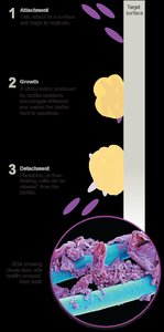
Biofilms are structured communities of microbes attached to surfaces and embedded in a self-produced matrix. They are highly resistant to antimicrobials and immune responses.
Biofilms can form on medical devices, teeth, water systems, and more.
Estimated 60–80% of human infections involve biofilms.
Laboratory Techniques in Microbiology
Culturing Microbes
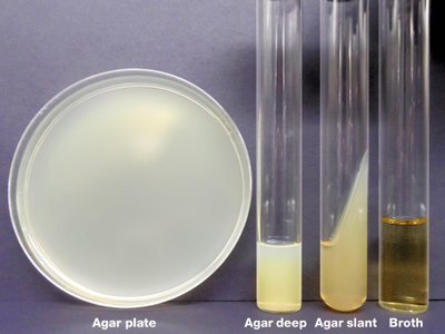
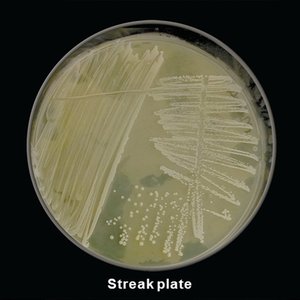
Microbes are grown in the laboratory using nutrient-rich media. Aseptic technique is essential to prevent contamination and ensure pure cultures.
Growth media: Nutrient broth (liquid) or agar (solid).
Pure culture: Contains only one microbial species.
Mixed culture: Contains multiple species.
Aseptic technique: Includes sterilizing instruments, decontaminating surfaces, and using protective equipment.
Staining Techniques
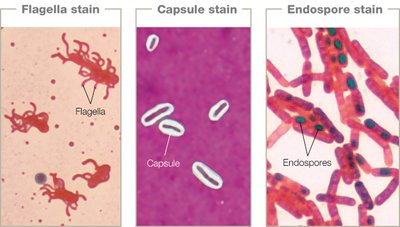
Staining increases contrast for microscopic observation. There are several types of stains:
Simple stains: Use one dye to reveal cell shape and arrangement.
Structural stains: Highlight specific structures (e.g., flagella, capsules, endospores).
Differential stains: Distinguish between types of cells (e.g., Gram stain, acid-fast stain).
Gram Stain
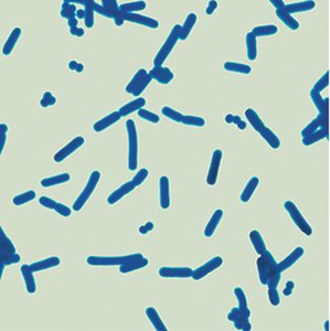
The Gram stain differentiates bacteria based on cell wall structure:
Gram-positive: Thick peptidoglycan wall, stains purple.
Gram-negative: Thin peptidoglycan, outer membrane, stains pink.
Acid-Fast Stain
Used to identify bacteria with waxy cell walls (e.g., Mycobacterium species). Acid-fast bacteria retain the red dye after acid wash; non-acid-fast bacteria do not.
Microscopy in Microbiology
Principles of Microscopy
Microscopy is essential for visualizing microbes. Key concepts include:
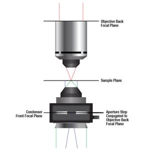
Resolution: Ability to distinguish two points as separate.
Refraction: Bending of light as it passes through different media.
Magnification: Enlargement of the image.
Numerical aperture: Light-gathering ability of the lens.
Types of Microscopy
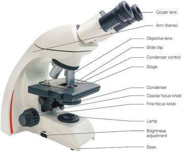
Different microscopy techniques are used based on the sample and desired information:
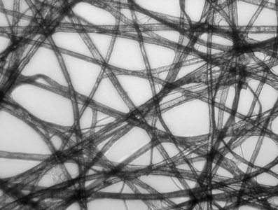
Light microscopy: Uses visible light; includes brightfield, dark field, and phase contrast.
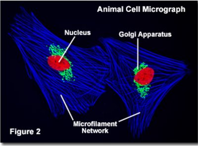
Fluorescence microscopy: Uses UV light to excite fluorescent dyes.
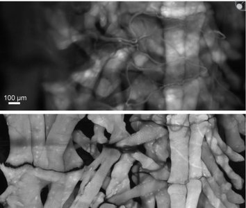
Confocal microscopy: Computer-assisted, produces 3D images.
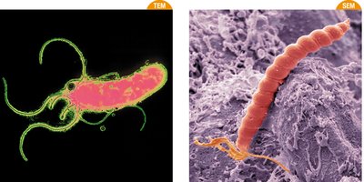
Electron microscopy: Uses electron beams for high resolution; includes transmission (TEM) and scanning (SEM) electron microscopy.
Sample Preparation for Microscopy
Microbial specimens can be prepared as wet mounts (live cells) or fixed mounts (killed and stained cells). Each method has advantages and disadvantages for observation and safety.
Wet mount: Observe live cells and motility; pathogens remain alive.
Fixed mount: Kills cells, preserves structure, safer for pathogens.
