 Indietro
Introduction to Prokaryotic Cells: Structure, Function, and Clinical Relevance
Guida di studio - Note intelligenti
 Appunti personalizzati basati sui tuoi materiali, ampliati con definizioni chiave, esempi e contesto.
Introduction to Prokaryotic Cells
Overview of Prokaryotic Domains
Prokaryotic cells are fundamental to life on Earth, having originated approximately 3.8 billion years ago. They are classified into two distinct domains: Bacteria and Archaea. Both domains share the characteristic of lacking a membrane-bound nucleus and organelles, but differ in their cell wall composition and membrane structure.
Bacteria: Possess peptidoglycan in their cell walls; fatty acids in membranes are linear.
Archaea: Possess pseudopeptidoglycan; membrane fatty acids are long-branched, and some form lipid monolayers for extreme environments.
Similarity: Both are unicellular and lack membrane-bound organelles.
Difference: Cell wall and membrane composition.
Basic Description and Size of Prokaryotic Cells
Prokaryotic cells are typically small, ranging from less than 1 µm to several µm in diameter. Their small size is due to reliance on diffusion for nutrient uptake, which is limited by the surface area-to-volume ratio.
Smallest: Mycoplasma species
Largest: Thiomargarita namibiensis
Shapes and Arrangements of Prokaryotes
Prokaryotes exhibit diverse shapes and arrangements, which are important for identification and pathogenicity.
Bacilli: Rod-shaped
Cocci: Spherical
Vibrio: Comma-shaped
Stella: Star-shaped
Coccobacilli: Ovoid
Spirochetes: Spiral-shaped, corkscrew motion
Monomorphic: One shape
Pleomorphic: Variable shapes, enhancing survival and transmission
Diplococci: Paired cocci
Streptococci: Chains of cocci
Staphylococci: Grapelike clusters
Diplobacilli: Paired bacilli
Streptobacilli: Chains of bacilli
Palisade: Clusters of bacilli
Example:
Streptococcus pyogenes, a Gram-positive coccus, forms chains (streptococci) and is clinically relevant in pharyngitis.
Binary Fission
Prokaryotic cells reproduce primarily by binary fission, a process that ensures rapid population growth.
DNA is copied
Cell grows
Chromosomes move to opposite ends
Septum forms at the midpoint
Two daughter cells are separated
Extracellular Structures
Plasma Membrane Structure and Function
The plasma membrane is a thin, flexible phospholipid bilayer acting as a selective barrier. Proteins embedded in the membrane serve as transporters, anchors, receptors, and enzymes, and are crucial for metabolic reactions such as ATP synthesis.
Selective permeability: Allows gases, water, and small noncharged substances to diffuse; larger or charged substances require transport proteins.
Membrane fluidity: Essential for function; affected by temperature and fatty acid composition.
Comparison of Bacterial and Archaeal Membranes
Bacteria: Linear fatty acids in phospholipids
Archaea: Long-branched fatty acids; some form monolayers for extreme heat
Cell Wall Structure and Function
Cell walls provide rigidity and protection. Bacteria use peptidoglycan, while archaea use pseudopeptidoglycan.
Gram Staining and Clinical Relevance
Gram staining differentiates bacteria based on cell wall structure:
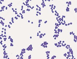
Gram-positive: Thick peptidoglycan, stains purple, lacks outer membrane
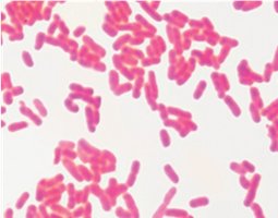
Gram-negative: Thin peptidoglycan, stains red/pink, has outer membrane with lipopolysaccharide
Gram-negative bacteria are harder to kill due to their outer membrane, which acts as a selective barrier.
Table: Comparing Gram-Negative and Gram-Positive Bacteria
Feature | Gram-Positive | Gram-Negative |
|---|---|---|
Peptidoglycan Layer | Thick (20–80 nm) | Thin (2–8 nm) |
Outer Membrane | Absent | Present |
Stain Color | Purple | Red/Pink |
Teichoic Acids | Present | Absent |
Lipopolysaccharide | Absent | Present |
Porins | Absent | Present |
Resistance to Chemicals | Lower | Higher |
Acid-Fast Staining
Acid-fast staining detects mycolic acid in cell walls. Acid-fast bacteria (e.g., Mycobacterium, Nocardia) appear red/pink and are slow-growing due to their waxy cell wall, which also impedes drug penetration.
Mycoplasma and L-Forms
Mycoplasma: Lack cell wall, have sterol-enriched membrane, pleomorphic, often intracellular
L-forms: Bacteria that lost their cell wall, resistant to certain stresses, may cause persistent infections
Transport Mechanisms
Passive Transport
Diffusion: Movement from high to low concentration
Simple diffusion: Small, noncharged molecules
Facilitated diffusion: Uses channel or carrier proteins
Osmosis
Osmosis is the diffusion of water toward higher solute concentration.
Isotonic: No net water movement
Hypertonic: Cell loses water, plasmolysis
Hypotonic: Cell gains water, possible lysis if cell wall is damaged
Active Transport
Primary: Uses ATP
Secondary: Uses ion gradients (symport and antiport)
Phosphotransferase: Group translocation, transfers phosphate to substrate (e.g., glucose)
External Structures for Adhesion, Movement, and Protection
Flagella
Flagella are protein filaments used for motility. Gram-positive bacteria have two rings for anchoring, while Gram-negative have four. The run-and-tumble system allows directional movement in response to stimuli (chemotaxis, phototaxis, aerotaxis).
Monotrichous: Single flagellum
Lophotrichous: Tuft at one pole
Amphitrichous: Flagella at both poles
Peritrichous: Flagella all over
Periplasmic flagella (axial filaments) allow spirochetes to move in a corkscrew motion.
Fimbriae and Pili
Fimbriae: Short, bristle-like, adhesive, common in Gram-negative bacteria
Pili: Longer, fewer, used for adhesion, movement, and gene transfer (conjugation)
Glycocalyx
Slime layer: Unorganized, loosely associated
Capsule: Well-organized, tightly associated
Glycocalyx aids in adhesion, protection from desiccation, and resistance to antibiotics.
Intracellular Structures
Nucleoid
Prokaryotic DNA is organized in a single, circular chromosome located in the nucleoid region.
Ribosomes
Prokaryotic ribosomes (70S) consist of a 50S large and 30S small subunit. They synthesize proteins and support the endosymbiotic theory.
Cytoskeleton
Composed of protein filaments, the cytoskeleton provides structural support.
Inclusion Bodies
Inclusion bodies are storage granules for substances such as carbon-fixing enzymes (carboxysomes) and magnetic iron (magnetosomes).
Endospores
Endospores are dormant, highly resistant structures formed by certain bacteria (e.g., Bacillus, Clostridium, Clostridioides). They survive harsh conditions and pose challenges in healthcare due to their persistence.
Sporulation: DNA is copied, packaged, surrounded by resistant layers, and released as a spore.
Examples: Bacillus anthracis (anthrax), Clostridium tetani (tetanus), Clostridium botulinum (botulism), Clostridium perfringens (gas gangrene), Clostridioides difficile (severe diarrhea)
Clinical Case Example: Streptococcal Pharyngitis
A 5-year-old patient diagnosed with streptococcal pharyngitis caused by Streptococcus pyogenes (Gram-positive, nonmotile, encapsulated). Penicillin-family drugs are effective due to the presence of peptidoglycan. No endospores or acid-fast properties are expected. Cell structures present include capsule, cell wall, plasma membrane, fimbriae, and ribosomes; absent structures include flagella, endospores, and acid-fast cell wall.
Classification: S. pyogenes is in the domain Bacteria, not Archaea.
