 Indietro
Microbial Cell Structure and Function: A Comprehensive Study Guide
Guida di studio - Note intelligenti
 Appunti personalizzati basati sui tuoi materiali, ampliati con definizioni chiave, esempi e contesto.
Chapter 2: Microbial Cell Structure & Function
Introduction to Microbial Cell Structure
Microbial cells exhibit a remarkable diversity in structure and function, which underpins their adaptability and ecological success. Understanding the architecture of microbial cells is fundamental to microbiology, as it informs their physiology, pathogenicity, and response to environmental changes.
Prokaryotic Cell Morphology
Cell Shapes and Their Biological Significance
Prokaryotes display a variety of cell shapes, each adapted to specific ecological niches and functions. The main morphological types include:
Coccus (pl. cocci): Spherical or ovoid cells.
Rod (bacillus): Cylindrical-shaped cells.
Spirillum: Spiral-shaped cells.
Spirochetes: Flexible, helical cells.
Appendaged bacteria: Cells with stalks or hyphae.
Filamentous bacteria: Cells forming long filaments.
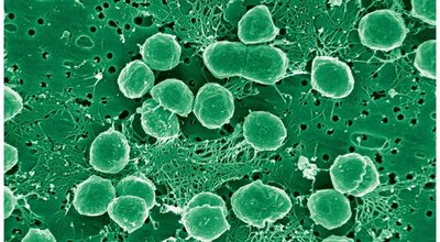
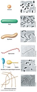
Example: Streptococcus species are cocci, while Escherichia coli is a rod-shaped bacterium.
The Advantage of Being Small
Cell size has profound implications for microbial physiology. Small cells have a higher surface area-to-volume ratio, which facilitates efficient nutrient uptake and waste elimination, supporting rapid growth and replication.
Surface Area (A):
Volume (V):
Surface Area-to-Volume Ratio: decreases as cell size increases.
Cells smaller than 1 µm may lack sufficient space for essential macromolecules.
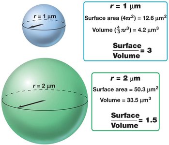
Example: Bacteria with a radius of 1 µm have a surface-to-volume ratio of 3, while those with a radius of 2 µm have a ratio of 1.5.
The Cell Wall: Structure, Function, and Clinical Relevance
Biological and Clinical Significance
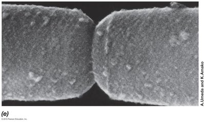
The cell wall is a defining feature of most prokaryotes, providing structural support, maintaining cell shape, and protecting against osmotic lysis. It is also a key determinant in host-pathogen interactions and a target for antibiotics.
Structural Support: Maintains cell shape and prevents bursting under osmotic stress.
Antigenic Properties: Surface molecules are recognized by the host immune system.
Antimicrobial Target: Many antibiotics (e.g., penicillin) and enzymes (e.g., lysozyme) disrupt cell wall synthesis or integrity.
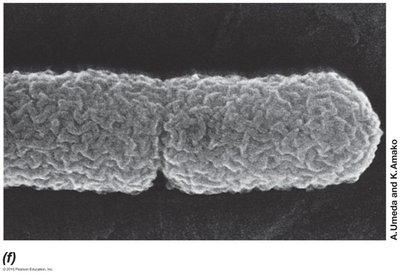
Gram-Positive vs. Gram-Negative Cell Walls
Bacteria are classified as Gram-positive or Gram-negative based on their cell wall structure and staining properties. This distinction is critical for diagnosis and treatment of bacterial infections.
Gram-Positive: Thick peptidoglycan layer, teichoic acids, no outer membrane.
Gram-Negative: Thin peptidoglycan layer, outer membrane containing lipopolysaccharide (LPS).
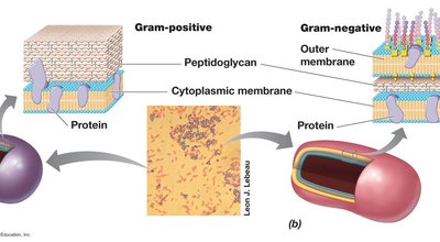
Gram-Positive Cell Wall Composition
Peptidoglycan: Thick, multilayered structure unique to bacteria.
Teichoic Acids: Polymers of glycerol or ribitol phosphate, confer negative charge and antigenicity.
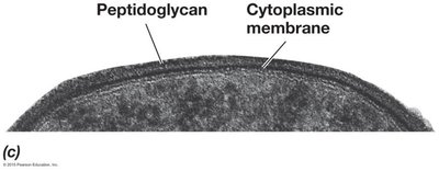
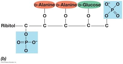
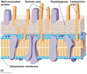
Peptidoglycan Layer: Structure and Function
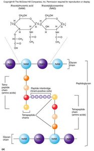
The peptidoglycan layer provides rigidity and shape to the bacterial cell wall. It is composed of glycan chains cross-linked by short peptides.
Glycan Chains: Alternating subunits of N-acetylglucosamine (NAG) and N-acetylmuramic acid (NAM).
Tetrapeptide Chains: Short peptides (often containing D-amino acids) cross-link glycan chains.
Gram-Negative Cell Wall Composition
Thin Peptidoglycan Layer: Located in the periplasmic space.
Outer Membrane: Contains lipopolysaccharide (LPS), proteins, and phospholipids.
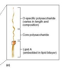
LPS: Composed of Lipid A (endotoxin), core polysaccharide, and O-antigen.
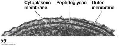
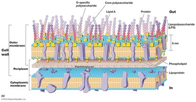
Lipopolysaccharide (LPS): Structure and Function
Lipid A: Anchors LPS to the outer membrane; toxic to humans (endotoxin).
Core Polysaccharide: Connects Lipid A to O-antigen.
O Side Chain (O-antigen): Highly variable; important for immune evasion.
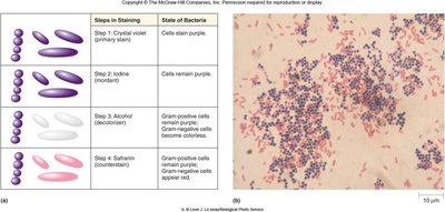
Gram Staining: Differential Staining of Bacteria
The Gram stain differentiates bacteria based on cell wall structure. The process involves staining with crystal violet, fixing with iodine, decolorizing with alcohol-acetone, and counterstaining with safranin.
Gram-Positive: Retain crystal violet-iodine complex due to thick peptidoglycan.
Gram-Negative: Lose crystal violet after alcohol-acetone treatment; stain pink with safranin.
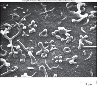
Bacteria Lacking Cell Walls
Some bacteria, such as Mycoplasma species, lack a rigid cell wall. They are resistant to antibiotics targeting peptidoglycan and rely on a unique plasma membrane for structural integrity.
Surface Layers External to the Cell Wall
Capsules and Slime Layers (Glycocalyx)
Many bacteria produce external polysaccharide layers known as capsules or slime layers, collectively termed the glycocalyx.
Adherence: Facilitates attachment to surfaces (e.g., dental plaque by Streptococcus).
Immune Evasion: Protects against phagocytosis (e.g., Streptococcus pneumoniae).
The Cytoplasmic (Plasma) Membrane
Structure and Function
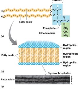
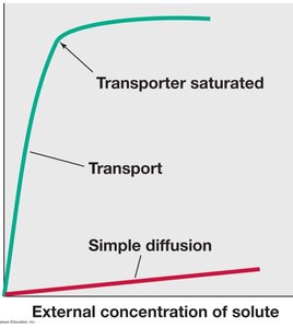
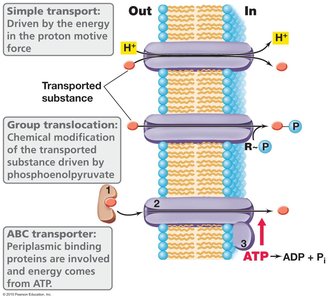
The cytoplasmic membrane is a selectively permeable barrier composed of a phospholipid bilayer with embedded proteins. It regulates the movement of substances into and out of the cell and is the site of energy generation.
Fluid Mosaic Model: Lipids and proteins move laterally within the membrane.
Transport Mechanisms: Include passive diffusion, facilitated diffusion, and active transport.
Motility and Surface Structures
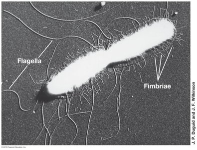
Flagella: Movement
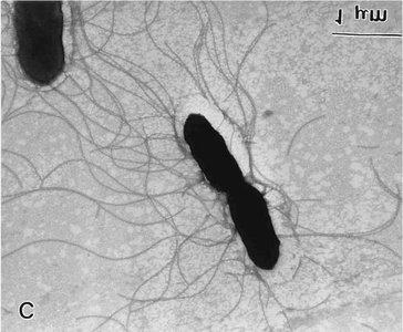
Flagella are whip-like appendages used for bacterial motility. Their arrangement and number vary among species and are adapted for movement toward or away from environmental stimuli (taxis).
Peritrichous: Flagella distributed over the entire cell surface.
Polar: Single flagellum at one end.
Lophotrichous: Multiple flagella at one end.
Types of Taxis: Chemotaxis (chemicals), phototaxis (light), aerotaxis (oxygen), magnetotaxis (magnetic fields), thermotaxis (temperature), hydrotaxis (water).
Pili and Fimbriae: Adhesion and Genetic Exchange
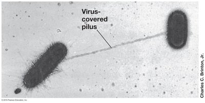
Pili are hair-like structures involved in adhesion and the transfer of genetic material (conjugation). Fimbriae are shorter and more numerous, primarily mediating attachment to surfaces.
Pilus: Used in conjugation (DNA transfer between cells).
Fimbriae: Mediate adherence to host tissues and surfaces.
Internal Structures
Bacterial Chromosome and Plasmids
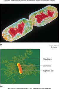
The genetic material of bacteria is typically a single, circular, double-stranded DNA molecule located in the nucleoid region. Plasmids are smaller, extrachromosomal DNA molecules that often carry advantageous genes, such as those for antibiotic resistance.
Ribosomes: Protein Synthesis
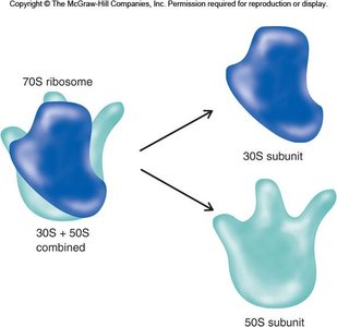
Bacterial ribosomes are the sites of protein synthesis. They are 70S in size, composed of 30S and 50S subunits, and are distinct from eukaryotic ribosomes, making them a target for certain antibiotics.
Storage Vesicles and Gas Vesicles
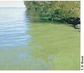
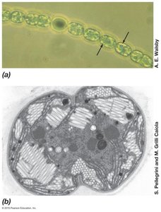
Bacteria may contain storage granules for nutrients (e.g., phosphate, glycogen) and gas vesicles for buoyancy, especially in aquatic environments.
Endospores: Survival Structures
Endospores are highly resistant, dormant structures formed by certain bacteria (e.g., Bacillus, Clostridium) to survive extreme conditions. They can persist for decades and germinate when conditions become favorable.
Resistant to: Heat, desiccation, UV radiation, chemicals.
Clinical Importance: Some endospore-formers cause serious diseases (e.g., anthrax, tetanus, botulism).
Summary Table: Comparison of Gram-Positive and Gram-Negative Cell Walls
Feature | Gram-Positive | Gram-Negative |
|---|---|---|
Peptidoglycan Layer | Thick | Thin |
Teichoic Acids | Present | Absent |
Outer Membrane | Absent | Present |
Lipopolysaccharide (LPS) | Absent | Present |
Sensitivity to Penicillin | High | Low |
Gram Stain Color | Purple | Pink/Red |
Additional info: This guide covers the essential aspects of microbial cell structure and function, focusing on prokaryotic diversity, cell wall architecture, and clinically relevant features. For further study, review the molecular mechanisms of transport, the role of surface structures in pathogenesis, and the unique adaptations of extremophiles.
