 Indietro
Microbial Cell Structure and Function: Study Notes for Chapter 2
Guida di studio - Note intelligenti
 Appunti personalizzati basati sui tuoi materiali, ampliati con definizioni chiave, esempi e contesto.
Microbial Cell Structure and Function
Introduction to Microbial Cell Structure
Microbial cells exhibit a variety of structural features that are essential for their survival, growth, and interaction with their environment. Understanding these features is fundamental in microbiology, as they determine the physiological capabilities and ecological roles of microorganisms.
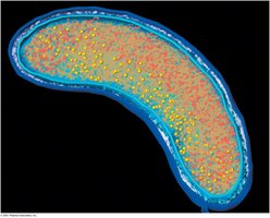
Cell Envelope: The cell envelope includes the cell membrane, cell wall, and, in some cases, additional layers such as capsules or S-layers.
Cell Membrane: The cytoplasmic membrane is a critical barrier and functional interface for transport, energy generation, and signaling.
Cell Wall: Provides structural support and protection; its composition varies among different microbial groups.
Naming and Classification of Microbes
Microbial taxonomy uses binomial nomenclature, which consists of a genus and a specific epithet to form the species name.
Example: Escherichia coli (E. coli) is a widely studied bacterium.
On first mention, the full name is used; subsequent mentions may abbreviate the genus (e.g., S. cerevisiae).
Strains are subtypes within a species, such as E. coli K12 used in laboratory settings.
Biosafety Levels in Microbiology
Microbiology laboratories are classified by biosafety levels (BSL) based on the risk posed by the organisms handled.
BSL1: Minimal risk, standard laboratory practices (e.g., E. coli K12).
BSL4: Maximum containment, for highly dangerous pathogens (e.g., Ebola virus).
Cell Membrane Structure and Function
Phospholipid Bilayer and Membrane Architecture
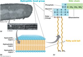
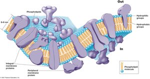
The cytoplasmic membrane is primarily composed of a phospholipid bilayer, which forms the fundamental permeability barrier of the cell.
Phospholipids: Consist of hydrophilic head groups and hydrophobic fatty acid tails.
Integral and Peripheral Proteins: Embedded or associated with the membrane, serving as transporters, receptors, and enzymes.
Archaeal Membrane Lipids
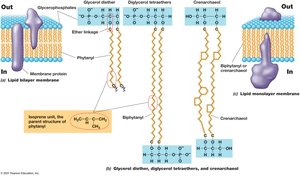
Archaea possess unique membrane lipids, including ether-linked isoprenoids, which confer stability under extreme conditions.
Glycerol Diether and Tetraether: Ether linkages instead of ester linkages found in bacteria and eukaryotes.
Monolayer Membranes: Some archaea have monolayer membranes for enhanced stability.
Functions of the Cytoplasmic Membrane
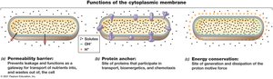
The cytoplasmic membrane serves as a permeability barrier, protein anchor, and site of energy conservation.
Permeability Barrier: Prevents leakage and controls entry/exit of substances.
Protein Anchor: Anchors proteins involved in transport, bioenergetics, and chemotaxis.
Energy Conservation: Generates and dissipates the proton motive force (pmf).
Proton Motive Force and ATP Production
The proton motive force (pmf) is generated by the electron transport chain (ETC) and is used to synthesize ATP.
ETC: Electrons are transported along membrane proteins, pumping protons out.
ATP Synthase: Protons return via ATP synthase, driving ATP formation.
Equation:
Transport Across the Membrane
Transport Systems
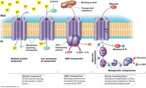
Microbial cells utilize various transport systems to import nutrients and export waste.
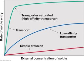
Simple Diffusion: Passive movement of solutes down their concentration gradient.
Facilitated Transport: Involves transporters that increase the rate of solute entry.
Active Transport: Requires energy, often via ATP hydrolysis or proton gradients.
Bacterial Cell Wall Structure
Gram-Positive and Gram-Negative Cell Walls
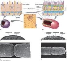
Bacterial cell walls are classified based on their response to the Gram stain, reflecting structural differences.
Gram-Positive: Thick peptidoglycan layer, retains crystal violet (purple).
Gram-Negative: Thin peptidoglycan layer, outer membrane, loses crystal violet and takes up safranin (pink).
Peptidoglycan Structure
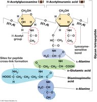
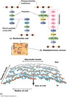
Peptidoglycan is a polymer of sugars and amino acids forming a mesh-like layer outside the plasma membrane.
Glycan Tetrapeptide: Repeating units of N-acetylglucosamine (NAG) and N-acetylmuramic acid (NAM) cross-linked by peptides.
Lysozyme: Enzyme that breaks down peptidoglycan.
Gram-Negative Cell Envelope
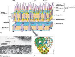
Gram-negative bacteria possess an outer membrane containing lipopolysaccharide (LPS), which provides additional protection.
Outer Membrane: Contains LPS, porins, and proteins.
Periplasm: Space between the outer and cytoplasmic membranes.
Gram-Positive Cell Wall
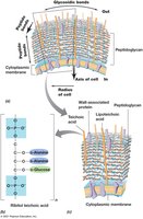
Gram-positive bacteria have a thick peptidoglycan layer with teichoic acids.
Teichoic Acids: Polymers that provide rigidity and regulate cell wall expansion.
Alternative Cell Envelope Structures
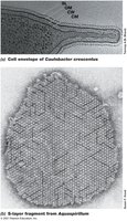
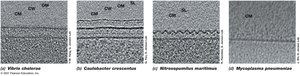
Some bacteria and archaea possess unique cell envelope structures, such as S-layers, acid-fast cell walls, or lack cell walls entirely.
Acid-Fast Cell Walls: Rich in mycolic acids, resistant to staining and antibiotics (e.g., Mycobacterium tuberculosis).
Cell-Wall-Deficient Bacteria: Mycoplasma and L-forms lack peptidoglycan, relying on membrane sterols.
S-Layers: Protein or glycoprotein arrays providing structural support.
Surface Structures and Storage Products
Capsules and Slime Layers
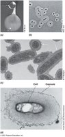
Capsules and slime layers are extracellular polysaccharide coatings that protect cells and aid in attachment.
Capsule: Well-organized, tightly bound layer.
Slime Layer: Loosely organized, easily washed off.
Fimbriae and Pili
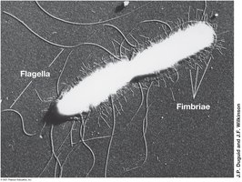
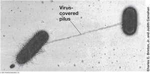
Fimbriae and pili are proteinaceous appendages involved in attachment and genetic exchange.
Fimbriae: Short, numerous, used for adhesion.
Pili: Longer, fewer, involved in conjugation and attachment.
Hami: Unique Archaeal Attachment Structures
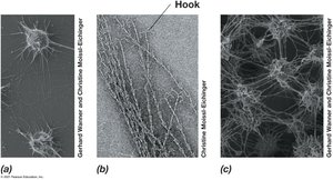
Hami are specialized appendages found in certain archaea, resembling grappling hooks for attachment.
Function: Enable strong adhesion to surfaces in extreme environments.
Storage Products
Microbial cells store nutrients in specialized granules or inclusions.
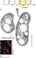
Poly-β-Hydroxyalkanoates (PHAs): Carbon and energy storage.
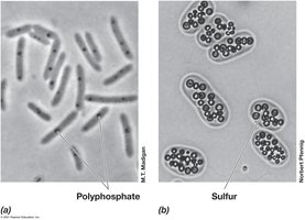
Polyphosphate and Sulfur Granules: Phosphate and sulfur storage.
Bacterial Endospores
Endospore Formation and Structure
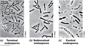
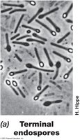
Endospores are highly resistant, dormant structures formed by certain bacteria (e.g., Bacillus, Clostridium) in response to adverse conditions.
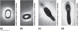
Structure: Includes exosporium, spore coat, cortex, inner membrane, and core containing DNA.
Function: Survival during extreme heat, desiccation, radiation, and chemicals.
Endospore Life Cycle
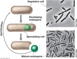
The life cycle involves vegetative growth, sporulation, and germination.
Sporulation: Triggered by nutrient depletion, involves asymmetric cell division and development of spore layers.
Germination: Occurs when conditions improve, returning to vegetative growth.
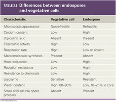
Differences Between Endospores and Vegetative Cells
Characteristic | Vegetative Cell | Endospore |
|---|---|---|
Microscopic appearance | Nonrefractile | Refractile |
Calcium content | Low | High |
Dipicolinic acid | Absent | Present |
Enzymatic activity | High | Low |
Respiration rate | High | Low or absent |
Macromolecular synthesis | Present | Absent |
Heat resistance | Low | High |
Radiation resistance | Low | High |
Resistance to chemicals | Low | High |
Lysozyme | Sensitive | Resistant |
Water content | High, 80-90% | Low, 10-25% in core |
Small acid-soluble spore proteins | Absent | Present |
Motility Structures
Flagella and Motility
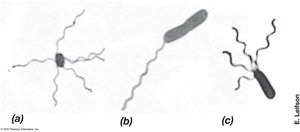
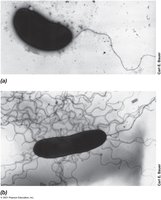
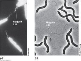
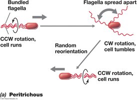
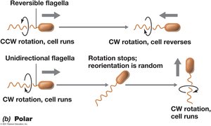
Flagella are whip-like appendages that enable bacterial motility. Their arrangement and mechanism differ among species.
Peritrichous: Multiple flagella distributed over the cell surface.
Polar: Flagella located at one or both ends of the cell.
Movement: Rotation of flagella propels the cell; direction and pattern depend on flagellar arrangement.
Summary
These notes provide a comprehensive overview of microbial cell structure and function, focusing on the diversity of cell envelopes, membrane architecture, transport systems, surface structures, storage products, endospore formation, and motility mechanisms. Understanding these features is essential for studying microbial physiology, taxonomy, and ecology.
